6 Study of fragmentary or burnt bones for establishing identity

1. Introduction:
Establishing of identity can be done, by taking into consideration four basic dimensions of the body namely; (i) sex of the person, (ii) age at death, (iii) height, and iv) ethnic identity. These four dimensions are approximated either in isolation or collectively, which provides a preliminary picture to be established, inorder to have a probable identification of the deceased individual.
Sex determination can be better manifested from the distinct recordable differences existing within the skeletons of both males and females. The more distinct, are the sexual differences, the more accurate would be the assignment of sex from the skeletal remains. In pre pubertal phase, sexual dimorphism is slight, hence sexual determination during the juvenile stage is difficult than in adult stage. The areas of high sexual dimorphism in adult are the skull and pelvis.
The complete identification of the deceased would be rather difficult if the skeleton is incomplete and fragmented. Thus, it is necessary that the remains should be carefully retrieved by the recovery personnel or the forensic archaeologists. Incases, where commingled remains or fragmented remains left from the explosion are found, the preliminary step is to reconstruct the body of the individual for assignation of skeletal remains. Moreover, it is extremely important to identify and assign a body to its correct side and in the correct position within a skeletal sequence.
Determination of sex:
Adults:
The foremost step of biological identification is sex determination, as the approximation of age and stature are sex dependent. For determination of the sex, to be reliable and well grounded, is based primarily upon whether the remains are complete and the level of sexual dimorphism inherent in the population to which the individual belongs. The male and female skeletons can be differentiated on the basis of two morphological differences. The male bones are generally larger and heavier due to the attachment of larger and more powerful muscle mass, larger size of the body and a period of delayed pubertal growth as compared to the female bones. Secondly, the male pelvis is primarily adapted to a bipedal striding gait, in contrast, the size and shape differences of the female reflect the biochemical compromise between efficient walking and the modifications necessary for the passage of large foetal head through the pelvis canal.
In regards to the shape of pelvis, the male pelvis is high and narrow whereas the female pelvis is wide and shallow, with a comparatively larger inlet and outlet dimensions. Another characteristic, which leads to the differentiation is the shape of greater sciatic notch, which is wide, shallow and big, incomparison to the ala of the sacrum compared to the width of its body incase of the females. Thus the most visible differentiation reflected posteriorly lies in the shape of greater sciatic notch, relative proportions of the body, alae of the sacrum and anteriorly in the sub-pubic angle, shape of the pubic bone.
In case of skull differentiation, the skull bones become more informative and visible at the time of puberty, where the skull of males are seen to have more muscle attachment whereas the skull of females tends to retain their more pedomorphic features. In case of males, features like glabella, supra orbital ridges, mastoid process and the nuchal and the malar areas become more prominent, as compared to the females, where all these features are smaller and the forehead remains vertical with more pronounced frontal and parietal eminences. As regards the orbits, in females they are more rounded, have sharper margins, and are larger relative to the upper facial skeleton.The mandible of males have a more prominent chin region, a more robust lower border, a greater body height, and a more prominent muscle markings. The angle formed between the body and ramus is more acute in the male than in the female.
Sex differences shows the relatively larger muscular development of the male, in rest of the post cranial skeleton, especially in relation to the overall joint size.
Juveniles:
As in the case of adults, sexual dimorphism can more precisely be observed through the pelvis and skull. But unlike as in the case of adults, sexual dimorphism with complete accuracy and reliability is difficult, incase of juvenile skeleton, as it does not reach a high level development which permits accuracy until pubertal modifications have taken place.
Age at Death:-
Chronological age estimation from the skeleton is based upon the measures of growth and development.
Growth is referred to as the increase in the size. This increase is size is accompanied by a change in form or function, which is development. Maturity is the attainment of adult form and function. The initial phase is primarily under the control of genetic and environmental factors which includes children and young adults who undergo changes that proceed in a relatively undocumented pattern at a moderately predictable rate. The changes related with age in an adult skeleton are quite prominent and significant, once maturity is reached. These changes are more individual and population specific, especially at the stage when maturity has reached and proceeds towards senescence and subsequent degeneration. The principle status of degenerative state includes factors like health status, occupation, nutrition, and endocrine function are principle status of degenerative status.
Juveniles:-
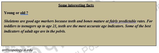
In the fetal and juvenile period, age estimation depends primarily on the appearance and fusion of the major centers of ossification and on the size of various skeletal elements, which includes growth and development. The dental age may be obtained from the time when eruption or mineralization begins and they are said to be much closer to the chronological age than the skeletal age. Dental age indicators have thus long been regarded as the gold standard as been less impacted by various influences such as sex, nutrition and ill health than bone growth and development.
Eruption is a continuous process by which teeth move from their crypts in the alveolar bone of the jaws to full occlusion in the mouth, often wrongly referred to as eruption. The presence of emerged teeth may be observed easily by the osteologist to give a rapid estimate of age, but increased accuracy can be obtained from evaluation from the calcification of the teeth. However, this necessitates radiographic analysis and comparison with defined stages of mineralization of both crowns and roots. This is a complex process that requires a considerable degree of experience and should be undertaken in conjunction with a forensic odontologist.
The skeletal age can be approximated from the size and aswell as the length of long bones of the body and from the developmental state of centres of ossification that has occurred. The accuracy of the age estimation is decreased postnatally, as the age advances due to the external factors that affects growth which consequently leads to individual as well as sex variation in stature, specifically during the adolescent growth spurt. The juvenile skeleton’s age can also be estimated from the developmental state of primary and secondary centres of ossification. The ossification in the embryonic and foetal stage initiates from the skull, vertebrae and primary centres of long bones and their girdles. The ossification centres starts in the embryonic centres starts with a non- descript spherical or in the form of ovoid nodules of bones, which remains unidentifiable in isolation and in skeletonized or commingled remains. Thus, in a forensic context, their use in the estimation of age would remain limited to the examination of body only, where a sufficient soft tissue remained to hold them in an anatomically identifiable position. This may ascertain its importance in legal cases, in forensic situations to identify and recognize if the foetus has reached full term and the presence of secondary centres of the distal femur, proximal tibia, calcaneus, talus is usually taken to signify this stage.When both primary and secondary centres of ossification comes to a point of recognizable distinctive morphology, estimation of age can be done. From the mid-foetal stage onwards, most of the bones of skull, vertebrae, ribs and major long bones of the limbs and girdles are in their recognizable stage, while others are recognizable only after the later childhood stage.
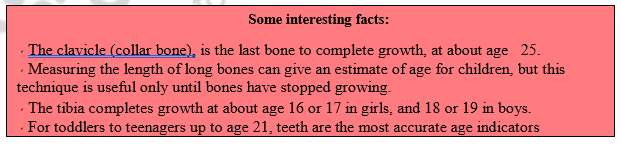
The bone elements which passess through the distinctive changes under a relatively short time period, their age estimation will be determined with greater accuracy. The fusion between the ischium and pubis at their point of ramii extends between 3-10 years, hence it is of limited value, whereas, the subsequent changes in the shape and the fusion between the main epiphysis and the tuberosity of the upper tibia takes place comparatively within a shorter time period and therefore are more useful and informative. The fusion between one or more primary and secondary centres of bone development is one of the most important phase, as it extends and covers a wide age range in response to the function of the soft tissues with which the bones are associated.
Young Adult Stage:
In the young adulthood stage, the estimation of age at death has much relevance as many areas of the skeleton remains incomplete till the second and third decades of life. This stage begins from the termination of height , which is marked by the fusion of all the long bones, till the time complete fusion of all the other epiphysis has reached. These include the regular jugular growth plate of the skull and post cranially, the sacral vertebral bodies, the iliac and ischial epiphyses, the ring epiphyses of vertebral bodies, the epiphyses of the scapula, costal notches of the sternum, and the medial epiphyses of the clavicle.
Adults:-
The process of aging utilizes majorly three areas of the skeleton i.e., the fusion of cranial sutures, morphological changes in the pubic symphyses, sacro iliac joint, and ventral aspect of the ribs.The epiphyseal morphology of the ventral demiface of the pubic symphyses undergo through an extended period of age-related changes upto 35-40 years. With the use of component phase analysis, this has been put to use for the estimation of age with each range, stating its own unique morphology. As a part of a multi-factorial method, the changes in the sacro- iliac have been used for the purpose of estimating age at death, but it was drawn out that the age ranges were too large for the application in forensics.
Sacro-iliac joint has more advantage over the pubic symphyses, since it being resistant to decay, but contrary in males, the surface of joint might not be visible after 50 years of age as it undergoes ankylosis. The examination of areas of hyaline cartilage ossification have proved to have some corroborative value in the prediction of age. The partially calcified tissue that might occur at this site, often not recovered, even if recognized can easily be damaged but ossification can arise at a surprisingly young age. Though occasionally, costal cartilage calcification had been reported in the later teenage years but it may not be visible until the middle of the fourth decade and in the upper four ribs until after the age of 50 years. Several studies have shown that a highly recognizable progressive pattern is visible with the increase of age, but timing is highly variable and the correlation between the actual age and the degree of ossification involves a wide margin of error.
The visible sign of degenerative diseases of the joint as marked by the lipping of vertebral bodies and osteophytes surrounding other joints and muscle attachments appears only after 40 years of age. The later decades of life are difficult to put to estimation with any degree of accuracy because these features are quite variable and are dependent on factors like genetics, nutrition, and lifestyle. With advancing years, the sclerotic fusion of manubriosternal joint increases, in the case of females. The primary cartilaginous joints between the manubrium and the costal cartilages of the first ribs may also synostose in older individuals but have never been reported in an individual where prior manubriosternal fusion has occurred. Piaget’s disease of bones, although only seen in 2-3% of adults, is rarely diagnosed in individuals less than 50 years of age. The synostosis of the sacro-iliac joint, seen after the 50 years of age is reported to be about 4 times more common in males so it could be used as corroboration of sex. Estimates of age at death is provided effectively as the continuous remodeling of the bones takes place throughout the adult life. Histomorphometric methods are based on the quantifiable patterns of intact and fragmentary osteons viewed in bone sections taken from specific sites in the body.
Stature:-
The approximation of stature from the skeleton of an individual can be simply obtained by calculating the stature by measuring the relative proportions of different parts of the body, both in relation to each other and in relation to the overall height of the individual. The stature can be estimated with greater accuracy, when the long bones of disarticulated adult skeleton. The greatest accuracy will be obtained when undamaged bones of unknown sex and ethnic identity are utilized, as the height of the individual is both sex and race dependent.

Ethnic Identity:-
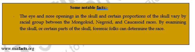
The determination of race or ethnic origin is the most difficult and unreliable attribute that the forensic osteologist must attempt to establish. The differences between the various ‘races’ of man are small, and the consequent variation exhibited in the skeleton is difficult to assess with any degree of accuracy.(St.Hoyme and Iscan,1987) It follows as the most obvious ethnic differences in the living are either displayed in the face or the integumentary system, so the skull is most studied skeletal element in this regard. Although, the history of anthropology is littered with explorers who have returned home with the skulls of many tribes to study, much of the earlier work concentrated on variation rather than on discrimination. Recently two computer based multivariate approaches have been developed on large database samples with the specific purpose of analyzing ancestry and therefore ethnic origin.
Ethnic identity from the limb bones utilizes variation in the inter-membral indices. For the upper limb, the brachial index is radial length × 100/ humeral length and for the lower limb, the crucial index is the tibial length×100/ femoral length .Both artificial dental work and innate dental traits can be useful for assigning ethnicity.
Positive Identification of Personal Identity:-
Once biological identity has been established, the forensic osteologist must attempt to personalize the information available so that the identity is achieved. During the Asian tsunami, the three principle means of personal identity were achieved through dental records, fingerprints and DNA analysis. All of these require a preexisting ante-mortem dataset be available for comparison. When it is known that a person is missing or deceased, then these records, should they exist, are passed to the existing authority. However, when an unidentified set of remains are found, then dental records are of no value, until a possible identity is achieved. Under these circumstances, the forensic osteologist must look to other possible evidence within the remains that may assist with the identification process.
Identification of a pre-existing medical condition or the presence of surgical hardware, may prove of value in establishing a productive line of enquiry. Equally, the forensic osteologist has a prominent role to play in the identification and reconstruction of trauma to the skeleton. (Black,2005).This may healed trauma that has occurred some considerable time before death or damage that could indicate the likely cause of death. Imaging techniques such as facial reconstruction, facial superimposition or facial art can be attempted with varying levels of success. These are speacialized areas that lie outside the capabilities of most forensic osteologists, and therefore it is essential that a good working relationship is developed between discipline-specific experts.
The forensic osteologist may feel pressured, by investigating authorities anxious to solve a case, to make a positive identification of an individual. It is essential to be absolutely sure that the remains are those of th e missing person and that the identification process rests on secure and verifiable evidence. The consequences could mean the end of distressing uncertainity and eventual closure for the family of a deceased individual with the acceptance of death and beginning process of grieving. In the case of possible crime, it could also mean the arrangement and trial of a suspect. Throughout the forensic examination of skeletal remains, there are various legal and sometimes political issues, and procedures, such as confidentiality and continuity of evidence that must be carefully followed. These usually follow cooperation with other personnel such forensic pathologists, archaeologists, odontologists and forensic examiners. The value of inter-disciplinary discussion and consultation cannot be over emphasized, and the forensic anthropologist must work as part of an active team rather than an isolated adjunct.
Summary:
When skeletal remains are discovered, it is the duty of the investigator to identify the decomposed or fragmented bones. Thus, the investigator begins his search with the answers to the following questions.
- Are the remains actually bones?
- Are the remains of human? It may be difficult to completely identify the fragmented bones, but in the hands of a skilled and experienced investigator, the investigator becomes easy.
- Is it a single bone or co-mingling of one or two bodies?
- What sex are the bones.
- What is the age of the person?
- What is the height (stature) of the person?
- What is the race or ethnic origin?
- Can a personal identity be discovered?
| you can view video on Study of fragmentary or burnt bones for establishing identity |