26 Body Fluids in Personal Identification- II

27.1 Introduction
Forensic serology is not to be confused with conventional serology, which deals solely with serum and its properties. Instead, forensic serology involves the identification of different types of body fluids. The identification of biological fluids during serology analysis is accomplished through presumptive and confirmatory testing. Presumptive testing refers to testing that is sensitive, fairly specific to the body fluid in question, and can be performed quickly. It allows an analyst to narrow down the number of items or areas of an item to focus on for further testing. Confirmatory testing is specific to the body fluid in question and sometimes also to a particular species. Confirmatory testing is still sensitive, but the time required for the testing can be much longer than that required for presumptive testing. In some instances, DNA analysis can be considered a type of confirmatory test because it is species, although not body fluid, specific for human DNA.
The types of evidence submitted to crime laboratories for serology/ DNA analysis are those items on which body fluids are thought to be present. A large majority of DNA/ serology cases involve sexual assaults. Evidence from these types of cases commonly includes sexual assault kits, complainant clothing, bedding, and, sometimes, suspect clothing. Items commonly submitted for testing include swabbing from crime scenes, clothing, weapons, or any number of other items that may possess bodily fluid. If an item is small, it can be submitted to the laboratory in its entirety. For larger items, stains can either be collected on a sterile cotton swab or a cutting from the item may be taken for submission at the lab. It is also possible to collect items that have been in contact with an individual’s mouth, such as cigarette butts, drinking cans, cups, bottles, gum, candy, toothbrushes, or ski masks. These items usually provide enough DNA for a profile to be established. Objects that have been touched or handled, such as a steering wheel, gun, phone, or even a fingerprint, may also contain biological evidence, which can be collected for analysis but may not always produce a DNA profile. Generally, these pieces of evidence do not contain a substantial amount of biological material and are processed for DNA without going through any type of serological screening to maximize the amount of sample available for DNA testing. Biological evidence is often left at burglary and theft scenes by the perpetrator. For example, a burglar may be injured breaking a window and leave blood at the crime scene, which can then be processed for DNA. In addition, DNA can be obtained from fingerprints or clothing left behind by a suspect. In cases of sexual assaults, seminal fluid examination is conducted to establish identity of the culprit. In some cases, when more than one accused is involved, vaginal swabs are also taken along with seminal fluid to prove that crime was committed by more than one person.
27.2 Learning Outcomes
In this module we shall be focussing on the importance of most of the body fluids such as semen, saliva, urine, feces, vomit etc. and their occurance at the scene of crime. We shall further learn to forensically examine them through different techniques. Students have already learnt about blood in module 26 therefore, it shall not be discussed here again. However, a brief outline of what we studied in module 26 body fluid in personal identification-1is given. We studied about what is blood, its constituents, preliminary and confirmatory tests. Species origin test and blood group determination. And finally the blood spatter analysis. In this module, body fluids in personal identification-2 students will learn how to test the presence of semen on a stained cloth or saliva from cigarette butt and likes and to extract DNA thereof.
27.3 Types of body fluids and their forensic examinations
The forensic significance of any body fluid lies in the fact that it may be eventually used to isolate blood group or DNA for personal identification or to narrow done the search for accused. But the real challenge is to screen the belongings and evidences to find the one possessing body fluid, so that it may be sent to laboratory for testing. Generally, a suspect’s body fluid on a complainant’s body or clothing, or a complainant’s body fluid present on clothing or items belonging to a suspect are the objects that hold the most evidentiary, or probative, value. For some cases, the most logical course of evidence examination is rather obvious. For example, in most cases of sexual assault, the identification of semen is central to supporting a claim of sexual assault. Furthermore, semen found on swabs in a sexual assault kit may have more probative value than semen found on clothing or bedding because, along with demonstrating the presence of semen on the complainant, semen can only survive inside a victim for a finite amount of time, whereas semen stains on clothing or bedding can have a much longer duration depending on whether the evidence is washed. For these cases, a determination can be readily made for the type of testing to perform and the most efficient order in which to process the items. Other cases are less obvious. If a sexual assault is oral, digital, or utilizing a foreign object, then it is useful to determine the details associated with the alleged assault to process the evidence most effectively. In these sexual assault cases, examining an item for the presence of semen may have no evidentiary value. All cases may be affected by any post-assault activity by the victim such as washing, wiping, eating, drinking, etc. The time between the assault and the examination can be a critical factor in the successful identification of body fluids because the longer the time span, the more evidence that may be lost. In any forensic case, the order of analysis for each test should be planned in advance to lessen the chance of losing evidence for the next test.
Homicide cases are more time-consuming to process than other types of cases because the victim cannot verbally relate any details of the assault. Homicides generally involve many items of evidence that must be analyzed because a determination cannot always be made regarding which evidence has the most value. Thorough crime scene investigation is essential to ensure that probative items in a case are collected and submitted to the laboratory. In cases such as these, communication with law enforcement is necessary to convey important case details to ensure that evidentiary items are processed in the most logical manner. When evidence is submitted, a determination must be made as to whether that evidence must go through serology screening or whether the evidence can be sent directly for DNA analysis. Generally, all evidence goes through serology screening first. However, cases involving samples with trace amounts of DNA may not benefit from serology screening. Paternity and remains identification cases also do not require any type of serology screening because only reference samples are processed.
Criminal paternity cases involve a sexual assault in which conception occurs. For these sexual assault cases, serology analysis is rarely performed. Instead, DNA analysis can be performed on the conceptus (living or aborted) and the alleged father to establish or disprove parentage (paternity testing).
The different types of body fluids that we shall discuss in this module are as follows:
i. Semen
ii. Saliva
iii. Urine
iv. Feces
v. Vomit
vi. Milk/ tears
And their forensic examinations and DNA isolation. It is important to note that most of the evidence processing and note taking occurs during serology analysis because this is usually the first time evidence is opened in the laboratory. Serologists are responsible for documenting the type, quantity, and packaging of the evidence received. In addition, a description of the evidence with notes and diagrams or pictures regarding the types of stains present and their location on each item is placed into the case file. Serologists also take detailed notes of their testing and outcomes, and this documentation is referenced during an analyst’s testimony during criminal proceedings. Thorough and precise note taking is essential because there may be a substantial amount of time between the completion of case analysis and an analyst’s testimony in court. It is also important in circumstances in which a different analyst must interpret the case notes.
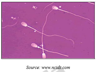
27.3.1 Semen
The identification of semen is important in many cases of alleged sexual assault. Semen is a body fluid produced by male individuals for fertilization. For forensic purposes, the composition of semen can be simplified into two components: seminal fluid and spermatozoa. Seminal fluid is a protein-rich body fluid originating primarily from the prostate and seminal vesicles. Spermatozoa, commonly referred to as “sperm,” are the male gametes, or sex cells, produced in the testis. Not all men produce spermatozoa. In men who have had a vasectomy, certain birth defects, or as the result of some diseases, seminal fluid will either not contain spermatozoa or contain very few. These are called as Oligospermins (with low sperm count) and Azoospermin (no sperm). Therefore, it is useful to be able to forensically test for the presence of both seminal fluid and spermatozoa. A healthy male usually ejaculate about 2-6 ml semen which has about 100 – 150 million sperm cells per ml. Its appearance is thick, yellowish white, opalescent, secretion having a characteristic odor known as seminal odor.

Presumptive Tests
i. Alternative Light Sources (ALS)
Under specialized lights, semen will fluoresce due to the presence of molecules such as Flavin and Choline-conjugated proteins. This colour will vary from blue to yellow depending on the light equipment used. This detection technique is highly presumptive because many molecules (natural and artificial) will fluoresce in a similar way as semen.
ii. Seminal Acid Phosphatase Test (SAP)
Also known as the Walker Test or Brentamine spot test. In the presence of Alpha-Naphthyl acid phosphate and Brentamine Fast Blue, AP will produce a dark purple color in less than a minute.
iii. Prostate Specific Antigen
Test detects prostate specific antigen (PSA). PSA is produced in high amounts by male prostate gland. This antigen can also be found in very small amounts of fecal material and sweat. Studies also indicate that PSA can exist in female urine and breast milk. Caution is urged when interpreting positive PSA results which are not confirmed by actual presence of sperm. This test is now obsolete.
iv. Choline Test
Stain extract is taken on a microscope slide and add a drop of Florence iodine. Place the cover slip and leave for 10 minutes. Brown coloured crystals of choline per iodide will be formed showing presence of semen in the stain.
v. Spermin Test
To prepare the extract- soak the stained cloth in 2.5% solution of trichloro acetic acid in attest tube and centrifuge for 1 hour. Take the supernatant and add equal volume of aq. solution of picric acid on a microscope slide. Yellow coloured obtuse or rhombic prism shaped crystals of spermin picrate will be formed showing positive for presence of human semen.
Confirmatory Tests
i. Christmas Tree Stain
Positive visual identification of sperm cells using a stain. Two main reagents are used consecutively to produce this distinctive stain: Picroindigocarmine stains the neck and tail portions of the sperm in green and blue, while the Nuclear Fast Red gives the sperm heads a read color and the tip of the heads a pink color.
Precautions: Sperm cells deteriorate quickly after ejaculation. Sperm survival will depend on the surrounding environment and type of surface. The sperm tails are the most susceptible to damage and will break down first. Therefore, the analyst must be trained to make visual distinctions between sperm heads and other types of cells in the mix.
ii. RSID (Rapid Stain IDentification) Test for Semen
It identifies the presence of the seminal vesicle-specific antigen, or semonogelin. This antigen is unique to human semen; therefore, there is no cross reactivity with other bodily fluids in males and females or with semen from other mammals. This test can also identify semen even if the stain was stored in less favourable conditions.
27.3.2 Saliva
The detection of saliva can be a useful tool in many types of criminal cases, although saliva testing is not requested as often as testing for semen or blood. While presumptive tests are available that can be used to indicate saliva, they have many limitations. Of the forensic laboratories that perform presumptive testing for saliva, the detection of amylase, an enzyme found at high levels in saliva, is currently the most widely utilized method. Amylase is found in a variety of body fluids but is more concentrated in saliva than in other body fluids. It should be noted that amylase is also found in plants and in some bacteria. In the body, amylase functions to break down starch into smaller molecules. A number of presumptive tests for amylase are available. While some of the presumptive tests are very sensitive for the presence of amylase, none can actually confirm the presence of salivary amylase. Therefore, many laboratories forego this test in cases where the quantity of saliva procured is very less. Instead, depending on the circumstances surrounding a case, some laboratories opt to save these samples for DNA testing.
Saliva is a colorless fluid secreted by 3 glands in the mouth. They are sublingual, sub-mandibular, and parotid. Saliva from parotid glands contains amylases, enzymes, which aid in the digestion of carbohydrates. Saliva is composed of 99% water and 1% includes electrolytes, enzymes, mucus. Humans produce 1-1.5 lts of saliva a day.
Presumptive Tests
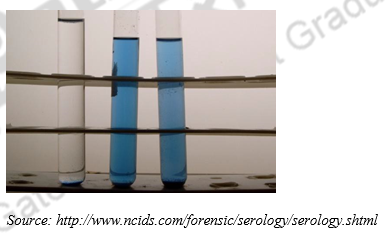
i. Phadebas Test
A chemical reagent called Phadebas is used to detect the enzymatic activity of the alpha-amylase enzyme, which is found in saliva. This enzyme is found in other organisms as well. Alpha-amylases from bacteria, fungi, or chimps are very similar in structure and function to that of the human alpha- amylase. Also, in humans, there are four variants of alpha-amylase, two of which are found in saliva, and the other two are secreted by the pancreas. This test is presumptive because it will give a positive result if the alpha-amylase enzyme from any organism is present.
- Place a small piece of the sample material in a 10 x 75 test tube. In a second tube, place an equal-sized piece of known saliva stain as a positive control. In a third tube add no sample (negative control).
- Add 1.0 ml distilled water and ¼ Phadebas tablet to each tube using clean forceps
- Vortex to mix thoroughly.
- A transparent dark blue supernatant of equal or greater intensity than the positive control is regarded as a positive test for amylase activity
- A blue color that is less intense than the positive control but darker than the negative control is considered inconclusive for presence of amylase. No blue color is considered negative for presence of amylase.
Amylase Test
Iodine solutions is added to suspected stain (saliva) and incubated at 37ᴼC followed by addition of starch solution. This causes starch to turn a deep blue color. But amylase is a starch hydrolyzing enzyme. The presence of amylase causes the disappearance of the blue color (due to hydrolysis of the starch) and can be used an indicator for the presence of amylase.
Confirmatory Tests
i. Starch Iodine Radial Diffusion Test
- Gel test plates (2% agarose, 0.1% soluble starch), Phosphate buffer, pH 6.9 10.0 ml, Agarose 0.2 g, Soluble starch 0.01 g is prepared by boiling and continue stirring constantly until all the agarose is dissolved. Divide gel solution and pour into 3-2″ disposable plastic Petri dishes. Allow to polymerize completely. Store gels inverted (to retard dehydration) at 4°C. Extract a small piece of stained material with 50 µl distilled water
Note: run at least one positive control consisting of a known dilution of fresh liquid saliva (1/500 in distilled water) and a negative control consisting of distilled water
**some laboratories add a second positive control consisting of a 1/100 dilution of fresh saliva
- Punch holes in gel plate with a vacuum pipette, leaving 1.5 cm between the sample wells.
- Place samples to be tested in the sample wells using a pipette. Each well holds approximately 4 µl of liquid.
- Cover the Petri dish and place in an incubator at 37°C for 6 hours or overnight.
- Stain the plate by pouring a 1:50 dilution of saturated iodine solution onto the surface. Rinse with distilled water
- Clear circles around the wells indicate areas of amylase activity. The diameter of the clear circle is proportional to the square root of the concentration of amylase. Record the diameter and results in notes.
ii. Phadebas Test and RSID Test for Human Saliva
The RSID Test for human saliva detects the alpha-amylase molecule itself, and specifically, the alpha- amylase from human saliva (in comparison to the testing for enzymatic activity as seen in the Phadebas test). Performing both of these tests is considered a confirmatory test.
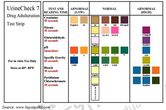
27.3.3 Urine
Urine is a transparent solution that can range from colorless to amber but is usually a pale yellow. In the urine of a healthy individual, the color comes primarily from the presence of urobilin. Urobilin in turn is a final waste product resulting from the breakdown of heme from hemoglobin during the destruction of aging blood cells. The smell of urine is pungent and strong and can be affected by the consumption of food. The pH of urine normally varies between 4.4 and 8 but is close to neutral- 7. In persons with hyperuricosuria, acidic urine can contribute to the formation of stones of uric acid in the kidneys, ureters, or bladder.
Urine is mostly tested in cases of alleged poisoning or administration of over dosage of drugs.

Tests for Urine
i. Creatinine Test
Reagent preparation: To 10ml sodium hydroxide(10%) add about 50 ml saturated solution of picric acid. Take extract on a slide and add few drops of reagent mixture. The intense red colouration is an indication of presence of urine.
ii. Urea Nitrate Test
Take the extract in test tube and add 2 drops of acetone to it. Now with a glass rod add a small drop of nitric acid into it. Positive result for urine shows formation of urea nitrate crystal at the junction.
27.3.4 Fecal Matter
Fecal Matter is excretory product of animal metabolism. It consists of undigested vegetable refuge, bilirubin, muscle fibers and cellular material. Its identification may be sometimes of great importance especially in cases of sodomy. For identification following examinations may be carried out. Fecal Matter is generally brown in color due to urobilinogen, in infants it is yellow due to unchanged bilirubin and milk diet. It has characteristic bad odour.
i. Microscopic Examination
Suspected stains are softened with distilled water, for about half an hour. A small amount of scraping from the stain is transferred onto a microscopic glass slide and a drop of Lugol’s iodine is added to it. The material is then covered with a cover slip and examined under microscope for the detection of undigested food particles, vegetable residues and muscle fibers.
ii. Urobilinogen Test
Reagent Preparation
Solution 1: 40% Alcoholic mercuric chloride solution.
Mercuric chloride 4 g Methanol 10 ml
Mix and store in stoppered bottle.
Solution 2: 40% Alcoholic zinc chloride solution
Zinc chloride 4 g
Methanol 10 ml
Mix and store in stoppered bottle.
Solution 3: Amyl alcohol.
Test: Urobilinogen is formed in the intestine by reduction of bilirubin. Urobilinogen is oxidized to Urobilin, which is soluble in alcohol. This test relies on the formation of a green fluorescent zinc- urobilin complex formed in the presence of neutral alcohol zinc salt.
Standard/ Control: A known fecal stain stained and unstained control should be tested each time the testing is performed. Use distilled water as a negative control.
Note: The species of origin of fecal matter, however, is not detected normally, unless it is contaminated with blood or mucus.
27.3.4 Vomit
The examination of vomit stains may prove or disapprove a suspect or victim’s alibi, support witness testimony and help in crime scene reconstruction. The transfer of vomit from one individual to another cannot be overlooked in crime cases of poisoning, drug overdose etc. in mostly suicidal attempts made by victims or in accidental poisoning cases. In such cases, usually stomach wash is given to the victim to vomit out all partially digested-undigested food items. Vomit may initially transfer from a victim to a suspect environment, i.e. clothing, dwelling or automobile. When stomach contents from a victim and foreign samples from the suspect environment are characterized and compared, similarities in food ingredients may suggest a common origin linking the suspect to the victim. The mere presence of vomit when none is reported in an alibi statement may be important to an investigator.
For the examination of vomit, presence of the following materials is to be taken into account:
1. Presence of mucus
2. Free HCl
3. Endothelial cells from gastric mucosa
4. Undigested and semi-digested food material
i. Test for Mucus
To the extract add 33% acetic acid drop by drop. Opalescence appears which may be due to mucus or lipoid substance or both. If on addition of more acetic acid opalescent does not disappear, presence of mucus is confirmed, because with excess of acetic acid lipoid globulins dissolve but not the mucus.
ii. Gunzberg’s Test (Test for Free HCl)
The reagent is prepared by 6 drops of 10% phloroglucinol in alcohol with 3 drops 10% vanillin in alcohol. In a porcelain evaporating dish one drop of suspected extract is placed and 1-2 drops of Gunzberg’s reagent is mixed at once. The contents are allowed to dry completely. A brilliant red color indicates free HCl.
iii. Endothelial Cells:
After centrifuging the extract for 10 minutes a thin film is made on a slide. The Endothelial Cells are observed under microscope.
27.3.6 Milk/ Tears
This is not very significantly important body fluid forensically. It is found present in lactating mothers. Generally, in cases of poisoning to kill infants or to prove the alleged pregnancy and illegal abortion of the infants are cases where it is examined. In almost all cases of drug abuse, drugs passes to some extent in breast milk, but the clinical significance of this depends upon:
i) The degree of drugs passage in to milk
ii) The amount of milk ingested by the infant at feeding
iii) Whether the infant absorbs the drugs
iv) Whether the drug affects the infant?
It is very difficult to determine that which drugs are contraindicated in lactating mothers, because of the very limited human studies on the subject. In assessing the impact of maternal medication on breast-feeding, the clinician must always weigh the many benefits of breastfeeding before start medication to nursing mothers also it is important to see if the medicines have any side effects on the lactating mothers, that may result in health hazards for the new born.
Same is the case with tears, it is also not important forensically usually because by the time the expert reach the scene of crime or by the time the exhibits reach the laboratory, tear etc. gets dried up leaving nothing behind for the expert to examine. Moreover, it does not constitute to prove that it is the result of a criminal act on;y.
27.4 DNA Analysis
Modern methods of collection and preservation of biological evidence for human identification by DNA Analysis are listed here. Investigators and laboratory personnel need to work together to determine the most probative pieces of evidence and to establish priorities. Given the sensitive nature of DNA evidence, officers should always contact their laboratory personnel or evidence collection technicians when collection questions arise. The high sensitivity of DNA determinations has even changed the way police investigators define biological evidence. Today, the sensitivity of PCR is so high that even as minute as 1 nanogram of DNA is sufficient to yield information to individualize evidence. With this technology in-hand, the horizon of the criminal investigator extends beyond the traditional dried blood or semen stain.
Note that, in practice, crime scenes samples may contain considerably less usable DNA depending on environmental conditions. DNA has been isolated from other sources, such as gastric fluids and fecal stains. However, it can be difficult to generate a DNA profile from these sources in case samples due to significant degradation. Several factors affect the ability to obtain a DNA profile. The sensitivity of PCR DNA typing methods is noteworthy, but still limited. The second concern is sample degradation. Prolonged exposure of even a large bloodstain to the environment or to bacterial contamination can degrade the DNA and render it unsuitable for further analysis. The third consideration is sample purity and DNA content of different types of samples.
What are some of the DNA technologies used in forensic investigations?
- Restriction Fragment Length Polymorphism (RFLP)
RFLP is a technique for analyzing the variable lengths of DNA fragments that result from digesting a DNA sample with a special kind of enzyme. This enzyme, a restriction endonuclease, cuts DNA at a specific sequence pattern know as a restriction endonuclease recognition site. The presence or absence of certain recognition sites in a DNA sample generates variable lengths of DNA fragments, which are separated using gel electrophoresis. They are then hybridized with DNA probes that bind to a complementary DNA sequence in the sample. RFLP was one of the first applications of DNA analysis to forensic investigation. With the development of newer, more efficient DNA-analysis techniques, RFLP is not used as much as it once was because it requires relatively large amounts of DNA. In addition, samples degraded by environmental factors, such as dirt or mold, do not work well with RFLP.
- PCR Analysis
Polymerase chain reaction (PCR) is used to make millions of exact copies of DNA from a biological sample. DNA amplification with PCR allows DNA analysis on biological samples as small as a few skin cells. With RFLP, DNA samples would have to be about the size of a quarter. The ability of PCR to amplify such tiny quantities of DNA enables even highly degraded samples to be analyzed. Great care, however, must be taken to prevent contamination with other biological materials during the identifying, collecting, and preserving of a sample.
- STR Analysis
Short tandem repeat (STR) technology is used to evaluate specific regions (loci) within nuclear DNA. Variability in STR regions can be used to distinguish one DNA profile from another. The Federal Bureau of Investigation (FBI) uses a standard set of 13 specific STR regions for CODIS. CODIS is a software program that operates local, state, and national databases of DNA profiles from convicted offenders, unsolved crime scene evidence, and missing persons. The odds that two individuals will have the same 13-loci DNA profile is about one in a billion.
- Mitochondrial DNA Analysis
Mitochondrial DNA analysis (mtDNA) can be used to examine the DNA from samples that cannot be analyzed by RFLP or STR. Nuclear DNA must be extracted from samples for use in RFLP, PCR, and STR; however, mtDNA analysis uses DNA extracted from another cellular organelle called a mitochondrion. While older biological samples that lack nucleated cellular material, such as hair, bones, and teeth, cannot be analyzed with STR and RFLP, they can be analyzed with mtDNA. In the investigation of cases that have gone unsolved for many years, mtDNA is extremely valuable. All mothers have the same mitochondrial DNA as their daughters. This is because the mitochondria of each new embryo comes from the mother’s egg cell. The father’s sperm contributes only nuclear DNA. Comparing the mtDNA profile of unidentified remains with the profile of a potential maternal relative can be an important technique in missing-person investigations.
- Y-Chromosome Analysis
The Y chromosome is passed directly from father to son, so analysis of genetic markers on the Y chromosome is especially useful for tracing relationships among males or for analyzing biological evidence involving multiple male contributors.
What are the preparatory steps taken before DNA analysis begins?
- Type of sample amount for DNA:
Liquid blood 20000-40000 ng/ml
Bloodstain 250-500 ng/cm
Liquid semen 150000-300000 ng/ml
Postcoital vaginal swab 10-3000 ng/swab
Plucked hair with root 1-750 ng/root
Shed hair with root 1-10 ng/root
Liquid saliva 1000-10000 ng/ml
Oral swab 100-1500 ng/swab
Urine 1-20 ng/ml
Bone 3-10 ng/mg
Tissue 50-500 ng/mg
- Evidence collection
All biological evidence is subject to deterioration. The careful collection and storage of this evidence will help ensure that this evidence is preserved so that useful information can be obtained from its analysis. Most DNA typing methods are robust, and dirt, grease, some dyes in fabrics, and other substances can seriously compromise the DNA typing process. Environmental insults will not change DNA allele “A” into allele “B”, but they can adversely affect the ability of the scientist to obtain a complete DNA profile from the sample. There are hundreds of varieties of physical evidence commonly submitted for examination to forensic science laboratories. Evidence that could be subjected to DNA analysis is generally limited to things that are biological in nature.
The following is a list of biological materials from which DNA has been successfully isolated and analyzed:
• Blood and bloodstains
• Semen and seminal stains
• Tissues and cells
• Bones and organs
• Hairs with follicles
• Urine and saliva (with nucleated cells)
Other types of biological evidence, such as tears, perspiration, serum and other body fluids without nucleated cells are not amenable to DNA analysis.
Blood, semen, body tissue, bone, hair, urine and saliva can be transferred to an individual’s body or clothing, or to an object or crime scene directly. Once liquid biological specimens have been deposited, they become stains and adhere to the surface or the substratum.
- Liquid Blood Specimens at Crime Scenes:
Liquid blood should be collected with a clean (preferably sterile) syringe or disposable pipette and transferred to a clean (preferably sterile) test tube. A blood clot can be transferred to a clean test tube with a clean spatula. A clean cotton cloth can be used to soak up liquid blood or a blood clot (avoiding areas containing only serum).
Wet blood samples, if they are collected, must be preserved in a suitable anticoagulant and kept in a refrigerator. These specimens should be submitted to the laboratory as soon as possible. Label the specimens with case number, item number, date, time, location, and evidence collector’s name.
- Wet Bloodstains: Small objects bearing wet bloodstains should be allowed to air dry, then collected as is. An effort should be made to preserve the integrity of any bloodstain patterns during packaging and transportation. Large objects that cannot be removed from a crime scene may have wet bloodstains on them. The wet blood should be transferred onto clean cotton cloth. Bloodstained cotton cloth must be allowed to air dry before packaging in a paper container. Each object and container must be properly labelled.
- Dried Bloodstains: on removable items dried bloodstains on weapons, garments and other movable objects should be collected separately by collecting the entire item. Each item should be placed in its own (paper) container, and these should be sealed and labelled properly. Dried bloodstains on solid, non-absorbent surfaces of immovable objects. The bloodstain pattern should be documented and sketched to the extent necessary. The stain can be tape lifted or scraped off the object onto a clean piece of paper. The tape lifter or the paper with blood crust can then placed into a “druggist fold”, and placed in an envelope which is sealed. Each item must be labelled properly.
- Semen and Seminal Stains: Document the semen evidence by notes, photography, videotape and sketchi Use a clean syringe or disposable pipette to transfer liquid semen to a clean, sterile test tube. Label the tube with the case and item number, date, time, location, and name of the collector. Keep the specimen refrigerated and submit to the laboratory as soon as possible. Alternatively, liquid semen can be transferred onto clean cotton cloth by absorption. The cloth is then air dried, packaged, sealed and labeled properly. Seminal stains on panties, clothing, bedsheets, pillows and other movable objects should be collected as is. If an article has a wet stain on it, the stain must be allowed to air dry thoroughly prior to collection of the article.
Collection of Urine, Saliva and Other Body Fluids follows the same rules as blood and blood stains.
- Issues concerning contamination
Because extremely small samples of DNA can be used as evidence, greater attention to contamination issues is necessary when identifying, collecting, and preserving DNA evidence. DNA evidence can be contaminated when DNA from another source gets mixed with DNA relevant to the case. There are some relatively simple steps that crime scene investigators can take in order to minimize the possible occurrence of contamination of biological evidence:
- Wear gloves. Change them often.
- Use disposable instruments or clean them thoroughly before and after handling each sample.
- Avoid touching the area where you believe DNA may exist.
- Avoid talking, sneezing, and coughing over evidence.
- Avoid touching your face, nose, and mouth when collecting and packaging evidence.
- Air-dry evidence thoroughly before packaging.
- Put evidence into new paper bags or envelopes, not into plastic bags. Do not use staples. Care should be taken to ensure that biological evidence is not contaminated during its
27.5 SUMMARY
In the present module we have learnt about most important body fluids encountered in most of the violent crime scene. We have learnt to distinguish and identify semen, saliva, urine, feces, vomit etc. using presumptive and confirmatory tests. Further we studied how one can extract DNA from them for personal identification. Students read about different techniques that can be used for DNA extraction such as RFLP, STR, PCR, mt DNA and Y-STR and their usability in different crime cases. For example mitochondrial DNA establishes the maternal lineage while Y-STR is passed on from father to son.
| you can view video on Body Fluids in Personal Identification- II |