25 Body Fluids in Personal Identification- I

26.1 Introduction
In the field of forensic science, serology and DNA analyses are closely related so much so that in many laboratories they are included within the same unit, collectively titled Forensic Biology. In the forensic laboratory, serology analysis refers to the screening of evidence for body fluids while DNA analysis refers to the efforts to individualize body fluids to a specific person. In most cases, body fluid identification is performed on evidentiary items before DNA analysis is attempted. Depending on the qualifications of laboratory personnel, analysts can be trained to perform either serology or DNA analysis or can be trained in both disciplines. While serology procedures have been employed for most of the twentieth century and the techniques have essentially remained unchanged, DNA has emerged in the forensic realm within the last two decades and its applications and technology are continuously developing.
26.2 Learning Outcomes
In this module we shall be focussing on the most important body fluid which is Blood and its examinations. Students will also learn about how to distinguish between any red coloured liquid and blood. Also whether the blood is of human origin or not. If the blood found at the scene is of human origin then blood group determination. Once established that the liquid is blood and it belongs to human origin and blood group is known, we will further do DNA analysis to ascertain the identity of the person whose blood we have examined. Finally, whether the blood spatter analysis yields any important information regarding the commissioning of act. We will also study the blood spatter and its importance at the scene of crime.
26.3 Blood
Blood identification is central to many homicide investigations and is also useful in cases involving aggravated assault, sexual assault, and burglary. The evaluation of blood evidence can be crucial to substantiate a complainant’s or suspect’s account of alleged events. The presence of blood on evidentiary items can be critical in establishing guilt or innocence during criminal proceedings. The analysis of blood evidence can be important not only in establishing which individual might have been bleeding, but also in the manner in which blood was deposited. Blood spatter interpretation can be valuable in determining how blood was deposited on an item or at a scene, thus making it useful in crime scene reconstruction.
26.3.1 Blood constituents
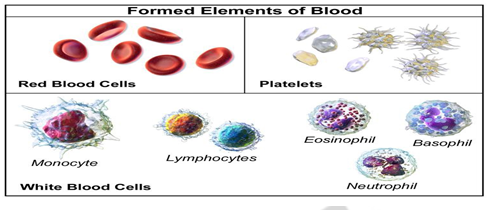
Blood accounts for 7% of the human body weight. The average adult has a blood volume of roughly 5 litres which is composed of plasma and cells. These blood cells consist of RBC (red blood cell or erythrocytes), WBC (white blood cells or leukocytes ) and Platelets or thrombocytes. By volume, the red blood cells constitute about 45% of whole blood, the plasma about 54.3%, and white cells about 0.7%.Whole blood contains red cells, white cells and platelets suspended in plasma. Plasma is the liquid portion of the blood that carries platelets, red cells and proteins throughout the body. Red cells contain hemoglobin, an iron-containing protein that carries oxygen throughout the body and gives blood its red color. White blood cells (WBCs), also called leukocytes, are an important part of the immune system. These cells help fight infections by attacking bacteria, viruses, and germs that invade the body. White blood cells originate in the bone marrow, but circulate throughout the bloodstream. Platelets are vital to life because they help prevent massive blood loss by helping your blood to clot.
26.3.2 Forensic examination of blood
One of the most common types of body fluid found at crime scenes, particularly the scenes of violent crimes, is blood. Though the appearance of blood is often quite distinct, chemical tests are essential to confirm its identity. Initially presumptive tests are used at the scene, which will merely confirm that the substance in question is most likely blood, though the species is not established at this point. Presumptive blood tests are usually based on the colour change or chemiluminescence of a particular reagent when it comes into contact with the haemoglobin in blood. The following are few commonly used presumptive tests for presence of blood.
Presumptive Tests
Presumptive tests are also known as preliminary tests, screening tests or field tests. These establish the possibility that a specific bodily fluid is present but do not conclusively prove the presence of a specific substance.
i. Phenolphthalein Test
It is also known as the Kastle Meyer Test. A Phenolphthalein solution is used to show the possible presence of blood based upon a peroxidase reaction of hemoglobin which produces a pink color.
ii. Benzidine Test
A benzene reagent is used to show the possible presence of blood based upon a peroxidise reaction of haemoglobin. A positive reaction in the presence of blood will give bluish green colour.
iii. Leucomalachite Green
Leucomalachite Green, is similar to the Kastle-Meyer test, replacing the phenolphthalein with leucomalachite green. When added to the substance, a green colour will be produced if blood is present.
iv. Luminol Test
Luminol is used in solution or sprayed onto suspected surfaces. This gives a strong blue fluorescence when viewed with a UV light. The Luminol reacts with hematin, a substanced formed as bloodstains age, and produces a luminescence which is best observed in the dark.
Confirmatory Tests
The confirmatory tests are also called as conclusive tests. These are conducted after preliminary test results positive. The following confirmatory crystal tests are performed for blood.
i. Takayama Test
Takayama reagent is added drop wise on a microscopic glass slide and subjected to mild eating. Hemo-chromogen crystals form confirms the presence of blood. The crystals are observable under a microscope and look like salmon-pink rhomboid crystals.
ii. Teichmann Test
Teichmann reagent is added drop wise in the blood stain on a microscopic slide and crystals are viewed under microscope. The reaction first converts the haemoglobin to hemin, and then the halides react with the hemin to form characteristic brownish-yellow rhomboid crystals.
Once it is ascertained that the given liquid is blood then we would test for specie origin. There are number of specie origin tests available. Some of which are listed below.
26.3.3 Specie Origin Tests
The specie origin test is done to establish whether the given blood sample belongs to a human being or not. If it does not belong to a human we do not do any further examination but if it is found to be that of a human then we will further do blood grouping and finally DNA analysis to pinpoint one person (or accused in forensic perspective). The species origin test is based upon antigen-anti body agglutination reaction. The antibodies are very specific to particular antigen only to which it binds.
i. Precipitin test
The precipitin test or ring test is conducted to determine the origin of species. In a test tube, 1ml of human antiserum is taken and few drops of suspected blood is added on top of it. If the blood is from human origin it will form a precipitin ring at the junction of two liquids to give positive result.
ii. Radial diffusion method
Most species identification uses radial diffusion of antigen and antibody. Here, antibody is mixed with agar gel on a plate into which punched crater are made (3mm -5mm) in which different antigens are taken. If a ring shaped precipitin band is formed around the crater from where the antigens migrate concentrically to react with the specific antibody, we would know that the blood taken in that crater is specific to the anti human anti serum present in gel o n plate. After the antigen diffuses for a time usually 2 -10 hours the migration ceases as the amount of antigen introduced exceeds the antibody in gel. This test is useful when we get different blood samples from crime scene and we do not know their origin.
iii. Double diffusion or Ouchterlony test
In the Ouchterlony test (also called as double diffusion in two dimension electrophoresis) an electric field is created rather than diffusion to move the blood extract (antigen) and antibody through the gel. Ouchterlony plates can be purchased or made in the laboratory. Extracts are made from stained areas of interest, and from nearby unstained areas (substrate controls). Note that the use of unstained controls is a fundamental principle in forensic immunologic testing. Stain and controls samples are loaded in the outer wells and a drop of anti-human antiserum is loaded into the center well. The process is repeated for antisera to other species, such as dog, cat, and cow; this may include the species from which the antiserum was obtained (e.g., rabbit).
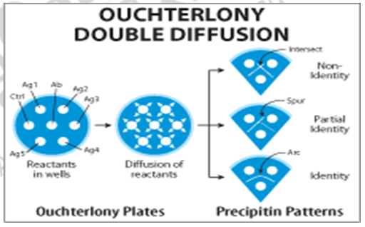
The plates are left at 4°C for a suitable period (which can range from a few hours to overnight) and the serum proteins and antibody molecules diffuse outward from the wells. A precipitin band is formed when the diffusing stain contains proteins that are recognized by IgG molecules in the diffusing antiserum. The precipitin band is sometimes clearly visible to the naked eye, but it is normal to stain the plates with amido black or other general protein stain, to enhance sensitivity and clarity.
iv. Cross-Over electrophoresis
Cross-over electrophoresis for species identification is conducted using agar at a pH of 8.6. Stain extracts are loaded into wells arranged in a line at the cathode end of the plate and the antiserum is loaded into wells at the anode end. During electrophoresis, the electric field drives the serum proteins towards the anode, but the IgG molecules, which are essentially neutral at this pH, are driven to the cathode by the process of electroendosmosis. The antigen-antibody precipitation occurs at the interface between the two rows of wells. Electroendosmosis occurs because the supporting medium acquires a net negative charge. If free, the negatively charged molecules would migrate to the anode, but this is not possible because the agar is immobilized on the plate. Instead, the effect is countered by positively charged water molecules migrating to the cathode.
26.3.4 Blood Grouping
a. ABO blood typing
Prior to the advent of DNA analysis for forensic science, other methods were developed for the comparison of biological fluid stains to individuals. The most common of these is ABO blood group typing. ABO blood typing identifies specific antigens present on the surface of blood cells. Within the population, individuals may have different forms of these antigens producing what is commonly referred to as a person’s “blood type.” Comparing the blood type obtained from an evidence stain to that of a known individual allows for the determination of whether the individual could have contributed to the stain.
A proportion of individuals known as “secretors” produce similar substances in other body fluids in addition to blood, which enables ABO typing to be performed on all body fluids in such individuals. The main drawback to ABO blood typing is that there are relatively few different ABO blood types throughout the population, making it difficult to individualize crime stains.
Nearly 40% of the population has blood type A and another 40% type O. In addition to being much less informative than DNA analysis, ABO typing requires a fairly large amount of sample for accurate testing, much more than is required for current DNA testing procedures. With the development of faster and more accurate DNA methods, most forensic laboratories have given up ABO testing. The ABO blood group system is widely credited to have been discovered by the Austrian scientist Karl Landsteiner, who identified the O, A, and B blood types in 1900.
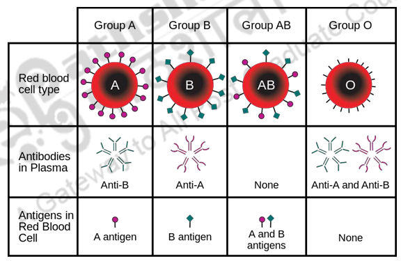
The basic principle of the ABO system is that antigens physically exposed on the exterior of red blood cells – differ between individuals, who have immunological tolerance only toward what occurs in their own bodies. Each antigen (also called as agglutinogen) binds with a specific antibody or agglutinins, which cause blood cells to clump and agglutinate if they belong to the specific related antigen. This way antigen A would agglutinate with only Anti-A antibody and the blood group would be A blood group. Similarly, antigen B would form precipitate with only anti-B antibody and the blood group will be called B blood group. While antigen A and antigen B if both react to give positive clotting with antisera A and B, the blood group would be AB. And in case where both antiserums A and B remain unreacted with given blood drop then the blood group would said to be O. Therefore, AB blood group people are universal acceptors because they have both antigens present and O blood group is universal donor. This reaction of antigen binding with specific antibody is useful against fighting infections, and may cause death when large amounts of opposite cells are encountered after a blood transfusion.
Rh Factor: In cases where antigen D is present we say blood group is Rh positive or (A+ or O+) whereas in cases where this antigen D is absent we call it as Rh negative or (B- or A-).
26.4 Blood Spatter Analysis
Bloodstain pattern analysis (BPA) is defined as the examination of the shapes, size, locations and distribution patterns of bloodstains, in order to provide an objective analysis of the physical events that gave rise to their origin by application of concepts of biology, biochemistry, physics and mathematics. Not only can these concepts help to define and reconstruct events associated in a bloodletting event but also may provide investigative leads/information. They can lead to new information, as well as supportive or non-supportive evidence for victims, suspect’s and witness statements. Besides this they give investors a better understanding in collection of relevant blood samples for DNA analysis. Bloodstain pattern analysis (BPA) is the interpretation of bloodstains at a crime scene in order to recreate the actions that caused the bloodshed. Analysts examine the size, shape, distribution and location of the bloodstains to form opinions about what did or did not happen. BPA uses principles of biology (behavior of blood), physics (cohesion, capillary action and velocity) and mathematics (geometry, distance, and angle) to assist investigators in answering questions such as:
- Where did the blood come from?
- What caused the wounds?
- From what direction was the victim wounded?
- How were the victim(s) and perpetrator(s) positioned?
- What movements were made after the bloodshed?
- How many potential perpetrators were present?
- Does the bloodstain evidence support or refute witness statements?
Because blood behaves according to certain scientific principles, trained bloodstain pattern analysts can examine the blood evidence left behind and draw conclusions as to how the blood may have been shed. From what may appear to be a random distribution of bloodstains at a crime scene, analysts can categorize the stains by gathering information from spatter patterns, transfers, voids and other marks that assist investigators in recreating the sequence of events that occurred after bloodshed. This form of physical evidence requires the analyst to recognize and interpret patterns to determine how those patterns were created. BPA provides information not only about what happened, but just as importantly, what could not have happened. This information can assist the investigator in reconstructing the crime, corroborating statements from witnesses, and including or excluding potential perpetrators from the investigation
26.4.1 Types of Stains
Bloodstains are classified into three basic types: passive stains, transfer stains and projected or impact stains.
a. Passive stains include drops, flows and pools, and typically result from gravity acting on an injured body.
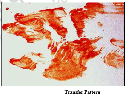
b. Transfer stains result from objects coming into contact with existing bloodstains and leaving wipes, swipes or pattern transfers behind such as a bloody shoe print or a smear from a body being dragged.
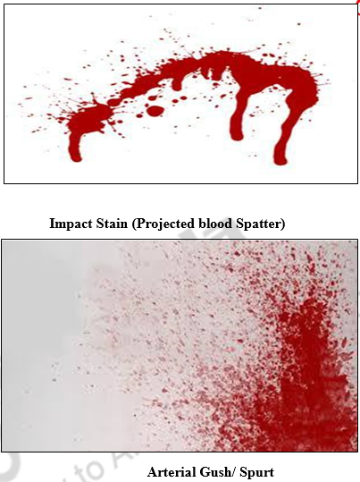
c. Impact stains result from blood projecting through the air and are usually seen as spatter, but may also include gushes, splashes and arterial splash.
Blood spatter is categorized as impact spatter (created when a force is applied to a liquid blood source) or projection spatter (caused by arterial spurting, expirated spray or spatter cast off an object). The characteristics of blood spatter depend on the speed at which the blood leaves the body and the type of force applied to the blood source.
Gunshot spatter includes both forward spatter from the exit wound and back spatter from the entrance wound. Gunshot spatter will vary depending on the caliber of the gun, where the victim is struck, whether the bullet exits the body, distance between the victim and the gun and location of the victim relative to walls, floors and objects. Typically, forward spatter is a fine mist and back spatter is larger and fewer
Cast-off pattern results when an object swung in an arc flings blood onto nearby surfaces. This occurs when an assailant swings the bloodstained object back before inflicting another blow. Analysts can tell the direction of the impacting object by the shape of the spatter (tails point in the direction of motion). Counting the arcs can also show the minimum number of blows delivered.
Arterial spurt refers to the spurt of blood released when a major artery is severed. The blood is propelled out of the breached blood vessel pumping of the heart and often forms an arcing pattern consisting of large, individual stains, with a new pattern created for each time the heart pumps.
Expirated spatter is usually caused by blood from an internal injury mixing with air from the lungs being expelled through the nose, mouth or an injury to the airways or lungs. Expirated spatter tends to form a very fine mist due to the pressure exerted by the lungs moving air out of the body. Small air bubbles in the drops of blood are typically found in this type of spatter
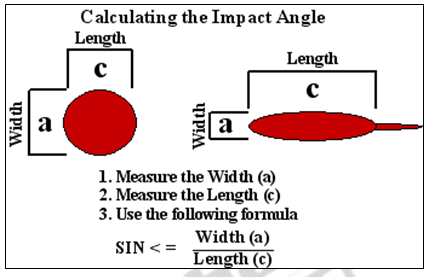
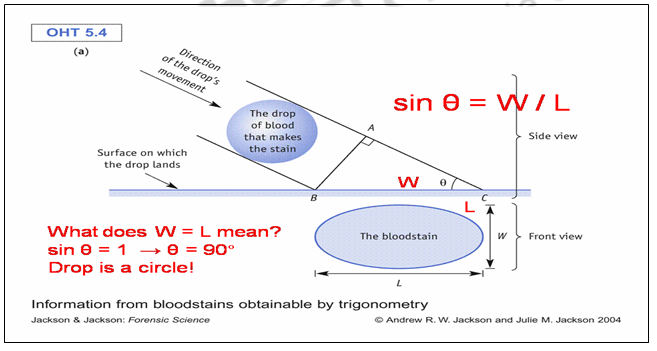
26.4.2 Angle of Impact
The length of the blood stain is the hypotenuse while the width is the opposite side of the angle of impact. The angle of impact can be calculated from the measurement of the opposite side which is the width and the length is hypotenuse of the blood stain.
Angle of impact = arcsin (opposite side/hypotenuse)
26.5 Summary
In the present module we have learnt about most important body fluid- blood encountered invariably in most of the violent crime scene. We have learnt to distinguish between any red coloured liquid and blood with illustrative presumptive and confirmatory tests. Further we studied how one can ascertain whether the given blood belongs to human or animal through various specie origin tests. Once established that the blood is of human origin we do ABO blood grouping tests to narrow done our suspects list. Lastly we understand the blood spatter analysis to study the angle of impact, weapon of offer.
| you can view video on Body Fluids in Personal Identification- I |