12 Determination of time of death through studies of bone, density method

Introduction
The estimation of age at death is an essential part in the reconstruction of population demographics and the individual analysis of human remains. Forensic anthropology is a speciality within physical anthropology. Forensic anthropologists work with skeleton to determine the identity of the deceased. They often work with forensic pathologists and forensic odontologist (dentists) to help determine the cause and manner of death and the postmortem interval (PMI) or length of time since death. Anthropologists can often tell the difference between an ancient and modern skeleton by the context of deposition. This includes the location, position, and condition of the body, and gives clues to the events which led to its burial.
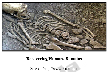
- Recover human remains
- Identity human remains
- Determine time or cause of death
Associated material evidence can also suggest the origin of a set of remains. Through the study of bones, an array of information can be ascertained regarding the remains including, but by no means limited to, age, gender, ethnicity, cause of death, and even indications of lifestyle such as where a person might have lived. The adult human skeleton consists of some 206 individual bones, with there being even more in the skeleton of a child, whose bones have not undergone certain fusion processes yet, and many of these bones may prove useful to the anthropologist.
Significance of Age
The estimation of age at death is an essential part in the reconstruction of population demographics and the individual analysis of human remains. After a person has reached adulthood (approximately 25 -30 years), bone have stopped growing. Changes to the bone are more subtle and there are fewer places on the skeleton where changes can be directly related to age. Other clues as to the age of skeletal remains may make an estimation of age possible. There are practical, criminal justice considerations out the age of bone. After a person has reached adulthood (approximately 25 -30 years), bone have stopped growing. Changes to the bone are more subtle and there are fewer places on the skeleton where changes can be directly related to age. This means that, when reliable knowledge about the age of bone remains, this must be taken into account when deciding if it is forensically significant.
Determining Time of Death
- Anthropological helpful if soft issue have decomposed
- If soft tissue is present, identification can be done by the pathologist
Stages of Decay
First, let’s look at the early stages of decay. The flesh of the body goes through five stages.
- The first stage consists of the ‘mortis‘phases. The blood isn’t being pumped through the body so due to gravity it pools in certain areas, and this is known as livor mortis.
- Shortly after this, the muscular tissue becomes rigid and incapable of relaxing, a state called rigor mortis.
- Next the body loses heat and cools in a process called algor mortis. Second, the body goes through bloat, in which means that microbes are rapidly growing and forming gases within the body. This is also usually when bugs and insects begin to feed and reproduce on the remains. In the third stage there is rapid loss of mass due to insect feeding and natural purging of fluids due to decomposition. Advanced decay is the fourth stage, and there is little left of the body at this point.Finally, the last stage is skeletonization when no flesh remains.
- Finally, the last stage is skeletonization when no flesh remains.
Age of Death
Although bones change throughout life in response to activity or inactivity, aging, disease, and injury, there are definite intervals during which bones are actively growing. Once they have reached maturity, the bones will not grow except for repairs and reaction to aging. Thus, the mechanisms by which the age at death is estimated are different for people who die while their bones are still growing (sub- adults) compared to those whose bones have stopped growing (adults).
- Dental eruption
- Bone size & maturity
- Epiphyseal closure
- Third molar root
- Sacral segments
- Pubic symphysis
- Cranial sutures
- Sternal rib end changes
- Peri-auricular margins
Age Estimation
- A number of methods are used to estimate the age-at-death of an individual from his or her skeletal remains
- The most common methods used to estimate the age of sub-adults (individuals under the age of 18) are bone formation and growth and dental formation and eruption
- Among the most common methods used to estimate the age of adults are cranial stature closure, changes to pelvic bones, and tooth wear
- It is easier to estimate the age-at-death of sub-adults because growth occurs in a known pattern
- Age estimation is not an exact science – age estimates are usually expressed in a range of years e.g. this individual was 40-50 years of age at the time of his death.
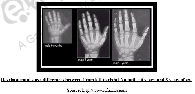
Using Bone development to determine age
Bones also have definite phases of growth that are age dependent. When the long bone starts to grow, they consist of the shaft or diaphysis and the end or epiphysis. As the individual develops these two fuses together at the growth area, called the metaphysis.

Within the metaphysis is the epiphyseal plate or growth plate, which lengthens as the immature bones grow. When the union is complete, growth ceases and what remains is a thin line at the growth area called the epiphyseal line. The union is not an event; it takes place over years. In general, union of individual bones takes place earlier for females than males. The clavicle in the shoulder has an epiphysis that fuses in women between the ages of 17 and 21 but in males the union take place between the age of 18 and 22.
Human Skull
There are some ossificatory processes of the immature skull which are useful in ascertaining the age of individual at death; though the use of suture closure to designate age has been challenged in recent years. This criticism is based on the evidence of high intra – group variability yet in a large number of cases this procedure is reliable.
- The frontal bone at birth consists of two halves which usually begin to unite in the second post – natal year. However, in certain proportion of cases this median frontal suture or metopic suture may remain open throughout life, thus demonstrating the phenomenon of metopism.
- The occipital bone at birth is in four parts: an upper part, two lateral portions and a basilar part. The upper portion (the squama) unites to the lateral portions by the end of the 5th year. The lateral parts join the basilar part before the 7th year. Later on the basilar part unites to the sphenoid bone by a strip of cartilage up to the 20th year. The closure of the basilar suture clearly demonstrates that the skull is that of an adult.
Look for the sagittal suture – the squiggly line that runs the length of the skull – and note whether is it’s completely fused. If it is, the remains are likely to be of someone older than 35. Look for a second line at the front of the skull — the coronal suture – which fully fuses by age 40.
- The suture at the back of the skull (lamboidal suture) begins closing at age 21, and by age 30 will have closed.
- By about age 32, the suture running across the top of the skull (sagittal suture), back to front, will have closed.
- By about age 50, the suture running side to side over the top of the skull, near the front (coronal suture), will have closed.
Look for the sagittal suture – the squiggly line that runs the length of the skull – and note whether is it’s completely fused. If it is, the remains are likely to be of someone older than 35. Look for a second line at thefront of the skull — the coronal suture – which fully fuses by age 40.
- The suture at the back of the skull (lamboidal suture) begins closing at age 21, and by age 30 will have closed.
- By about age 32, the suture running across the top of the skull (sagittal suture), back to front, will have closed.
- By about age 50, the suture running side to side over the top of the skull, near the front (coronal suture), will have closed.
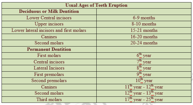
Dentition
The eruption of the temporary and permanent dentition is a much better guide to the age of the individual at death. The sequence of both deciduous and permanent tooth eruption is quite consistent because the teeth erupt in a stated order and at average ages for each tooth. This sequencing is substantially the same for both sexes and all races of modern man studied so far. As such sexual and racial variation in the age of the eruption of teeth may disregard in attempting to age specimens.
Vertebrae
- The neural arch joins the body or Centrum of each cervical vertebra at about three years of age, each thoracic and lumber vertebra at about 6-7 years.
- The epiphyses of the centra or annular epiphyses appear at puberty and generally fuse by 20 year.
- Union of the sacral vertebrae is completed after the 25th year.
- Degenerative pathologies e.g. spondylitis, arthritic symptoms and exostoses are likely to appear after 35 years of age.
Using pubic and ribs bones to determine age
The main areas in the body where age determinations are made in adults are the pubic bones and rib bones. In adults there are several ways of determining age at death. One of the most common methods is the microscopic observation of the condition of the pubic symphysis. The left and right hip (pelvic) bones join at the pelvis. Where these join there is a symphysis or space that has small amount of cartilage. When the cartilage is removed and the bones are separated, the shape and the surface texture on the medial (inner) surface can be examined. These portions of the pelvic bone change in a predictable way as a person ages. This surface is rough and billowed in younger adults, but by age 35 the surface become increasingly smoother and develops a rim. After age 35, the symphysis steadily degenerates and the surface beings to erode. The older the person at death, the more rough and uneven these bones will be. Forensic anthropologists will compare this against a database of standard markers to learn the age of the skeleton. Check if there are any soft marks on the cartilage which are left by childbirth as the bones soften to allow easier birth.
- The ramii of the ischium and pubis unite at about 7-9 years in males and 5-7 years in females.
- Union of all three segments in the acetabulam is usually complete by about 16 years of age.
In addition to the pubic symphysis, the ends of the ribs that meet in the front of the body (the sterna ends) also change as a person gets older. Examine where the ribs join the sternum. This is also a good indicator of age. A forensic anthropologist will compare it against a database of standard markers and it is often more accurate as it is not a weight-bearing bone and remains unaffected by childbirth.
Wrist
Examine the wrists, as bones often hold clues to the primary work of the decedent. Bony ridges form where the muscles were attached and pulled over the years. A forensic anthropologist might find a bony ridge on the wrist and decide the dead person may have been someone who used their hands for a living, such as a chef or seamstress
Long Bones
- The head of the femur fuses with the neck at about 14-15 years in females and 18-19 years in males.
- The head of the humerus fuses with the shaft at about 17 years in females and 19-20 years.
- The olecranon process of the ulna appears in both sexes around 11 years of age; fusion is complete in females around 15 years of age, while in males it is complete around 17 years.
The age order of the union of epiphyses has been determined accurately by Stevenson (1924) on the basis of large series of skeleton of known age. The sequence ids definite and consent for main irrespective of sex and race.

Thus it is clear that the estimation of chronological age at death draws upon a wider range of observation. However, it depends for its accuracy on the verification of sex on account of developmental precocity in the female. Besides, there is variation between populations in the skeletal manifestation of chronological age due to genetic as well as nutritional and other environmental differences.
Isotope Analysis
Bones and teeth are usually subjected to isotopic analysis in cases of dating skeletonised human remains, though if present hair and nails may also be used. Establishing the age of a set of remains, as in how long ago the individual died, is often a difficult task. With more recent sets of remains, there may still be some tissues present on the body to help pinpoint the age of the body. Certain soft tissues and ligaments can last for up to 5 years, so the presence of these may at least be able to narrow down time since death to the last few years. Typically, isotope analysis can prove to be particularly beneficial in establishing the likely age of remains. Among the most common elements to be studied in isotope analysis are carbon, nitrogen, oxygen, strontium and hydrogen. This branch of study, which can be focussed upon unstable or stable isotopes, is based on the principle that many elements within the body exist as various isotopes, many of which are taken into the body by eating, for instance.
Despite the benefits of this technique, it is not as accurate as would be ideal and establishing time since death using isotope analysis can only allow for an estimation. However it must be taken into account that exposure to certain conditions can affect the isotopes present in a sample and the ratios of these isotopes. Furthermore, isotopes are ubiquitous in the environment thus it is entirely possible for detected isotopes in a sample to actually be the result of contamination.
Summary
Bones are live and carry on all life functions. The condition of bones can tell investigators about a person’s health and nutrition during life. The identification of factors differentially expressed in fusing sutures has led to multiple targets for future research. The age of a person at death can be estimated by analysis of a number of bones. During life, many of the 450 bones a person has at birth grow together, finally forming 206 bones. As the cartilage between them is replaced, an epiphysis line is visible. When the cartilage is fully replaced, the line is no longer visible. This information can be used to approximate a skeleton’s age. When the skeleton is first discovered, take samples from around the remains including any bugs you come across. Insects such as blowflies have a very distinct lifecycle and often plant their eggs on newly deceased bodies. By identifying the stage of the lifecycle of bugs, a near-exact time of death can be established. This science is known as forensic entomology.
Finally it may be pointed out that the determination of age at death from the post cranial skeleton is quite difficult in mature individuals. However, the most reliable indicators are the face of the pubic symphysis, certain features of the scapula, closin
| you can view video on Determination of time of death through studies of bone, density method, Human skull |