13 Skull Suture Pattern – Individualization based on Suture Patterns

Introduction
Forensic anthropologists work with Human skull to determine the identity of the deceased. In present times of frequent air travel, accidents and mass disasters, the identification of individual from sutural pattern will be effective for setting insurance claims, showing certainty of death of a particular individual, and performance of last rites. In medico-legal cases and mass disasters, the identification of individual is critical; individualization may refer to discrimination or perception of the individual with a group or species. The bones and teeth of the craniofacial complex, key identification tools for the forensic odontologist, effectively distinguish one person from other and one population from another, and are used to determine the race, age and sex of a person.
Human Skull
The skull refers to the bones which constitute the skeleton of the head and face including the mandible. The term cranium is used to describe the skull without the mandible. The cranium is divided into an upper box like part the calvarium, which contains the brain and lower anterior part which forms the facial skeleton. The fact that the primary shape of the skull is genetically determined is well demonstrated by transplantation of embryonic skeletal tissues and organ culture studies. Other characteristics of the bone seem to be self determined. Mechanical influences may not be operative at the time of establishment of the primary form of the bone. Later on muscles become active and may influence bone growth even in the pre- natal life.
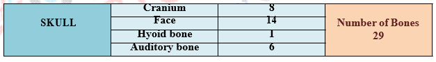
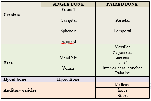
The skull consists of 29 bones which in the adult are firmly united with each other except for the mandible. The immovable joint in the skull are called sutures. The edges of bones forming these joints are frequently serrated so that they interlock. The brain is located inside the cranial vault, a space formed by bones of the skull and skull base. Everything inside the cranial vault is ‘intra-cranial’ and everything outside is ‘extra-cranial’.
Skull Bones
Bones of the skull and skull base – frontal, parietal, occipital, ethmoid,sphenoid and temporal bones – all ossify separately and gradually become united at the skull sutures. The skull has inner and outer tables of cortical bone with central cancellous bone called ‘diploe’.
A human skull is almost full sized at birth. However, the eight bones that make up the cranium are not yet fused together. This means that the skull can flex and deform during birth, making it easier to deliver a baby through the narrow birth canal. These individual plates of bone fuse together after about 24 months to form the adult form of skull.
Exterior of the Skull
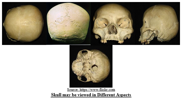
The skull may be viewed in different aspects from above, below, behind, the front and the sides. Accordingly they have been named as followed:
- Norma Verticalis (viewed from above)
- Norma Occipitalis (viewed from behind)
- Norma Frontalis (viewed from the front)
- Norma Lateralis (viewed from the side)
- Norma Basalis (viewed from the below)
Cranial Sutures
The main sutures of the skull are the coronal, sagittal, lambdoid and squamosal sutures. The metopic suture (or frontal suture) is variably present in adults.
- Coronal suture – unites the frontal bone with the parietal bones.
- Sagittal suture – unites the 2 parietal bones in the midline.
- Occipito-mastoid suture- unites the temporal bones with the occipital bone.
- Lambdoid suture – unites the parietal bones with the occipital bone.
- Squamous suture – unites the squamous portion of the temporal bone with the parietal bones
- Metopic suture – (if present) unites the 2 fontal bones.
- Parietal Mastoid suture- unites the parietal bones with the temporal bone.
Structure and Development of Human Skull
Human skull varies in their shape and gross appearance. Their shape and structure is affected by genetic, metabolic and mechanical factors. Each skull is the result of a long functional history through innumerable successive generation. Human skull is one of the hardest structures of the body; it also possesses a certain degree of toughness and elasticity.
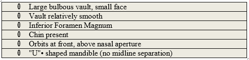
- Cranial morphology differs dramatically in humans due to the uniquely large brains that humans have compared to body mass. It is quite distinct because it has large, rounded braincase, and flat or orthognathic face in profile.
- “U” shaped singular construction of the human mandible.
- Human crania are oriented on a vertical axis and the orbits are located in front and above the nasal aperture. These orientations also cause the position of the foramen magnum to be located inferiorly in humans.
Skull Suture Development
The human Calvarium is formed primarily from five bones, the paired frontal and parietal bones and the unpaired interparietal bone. The cranial sutures allow for deformation of the skull during birth and subsequent growth of the skull. The frontal bones are separated along the midline by the metopic suture and the parietal bones are separated by the sagittal suture. The coronal suture separates the frontal and parietal bones and the lambdoid suture separates the parietal bones from the single occipital bone posteriorly. These bones are formed by intramembranous ossification and growth occurs perpendicular to the orientation of the suture. In normal development, the metopic suture undergoes fusion during the first year of life while other sutures fuse at various times during adulthood.
The plates of the membranous bones making up the Calvarium of the skull are each derived from the primary ossification centre, from which bone formation spreads outward. However, the individual plates do not fuse with each other during prenatal development. As a consequence, new born babies have unclosed (open) sutures and fontanelles. These temporary discontinuities between the bones of the Calvarium aid passage of the head through the birth canal at childbirth and permit an increase in the size of the skull to match brain growth after birth. The posterior (smaller) fontanelle closes during the first year, and the anterior (larger) fontanelle closes during the second year after birth. However, some of the sutures remain open until adulthood.
Determining Identity through Human Skull
The bones and teeth of the craniofacial complex, key identification tools for the forensic odontologist, effectively distinguish one person from other and one population from another and are used to determine the race, age and sex of a person.
Age Estimation of both the Living and Deceased
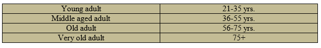
Human skull; clues as to the age of skeletal remains may make an estimation of age possible. There are practical, criminal justice considerations from the age of bone. The estimation of age at death is an essential part in the reconstruction of population demographics and the individual analysis of human remains. After a person has reached adulthood (approximately 25 -30 years), bones stop growing. Changes to the bone are more subtle and there are fewer places on the skeleton where changes can be directly related to age.
There are some ossificatory processes of the immature skull which are useful in ascertaining the age of individual at death; though the use of suture closure to designate age has been challenged in recent years. This criticism is based on the evidence of high intra – group variability, yet in a large number of cases this procedure is reliable.
- The frontal bone at birth consists of two halves which usually begins to unite in the second post – natal year. However, in certain proportion of cases this median frontal suture (or metopic suture) may remain open throughout life, thus demonstrating the phenomenon of metopism.
- The occipital bone at birth is in four parts: an upper part, two lateral portions and a basilar part. The upper portion (the squama) unites to the lateral portions by the end of the 5th year. The lateral parts join the basilar part before the 7th year. Later on, the basilar part unites to the sphenoid bone by a strip of cartilage up to the 20th year. The closure of the basilar suture clearly demonstrates that the skull is that of an adult.
An overview of milestone studies in suture development; according to Todd and Lyon each cranial suture presents two aspects:
- The Endocranial (seen on the inside of the skull) and
- The Ectocranial (seen on the outside of the skull).
Closure begins approximately at about the same time in both aspects. However, the Endocranial closure is more regular and reliable. It may be kept in mind that sometimes there may be ‘lapsed union’ in which bone heaps up along the edges of a suture without the closure of the suture taking place. Such ‘lapsed union’ may affect the whole of the suture or part of it. It is usually common in the sagittal and lambdoidal suture.
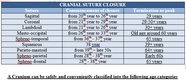
Look for the sagittal suture – the squiggly line that runs the length of the skull – and note whether is it’s completely fused. If it is, the remains are likely to be of someone older than 35. Look for a second line at the front of the skull — the coronal suture – which fully fuses by age 40.
- The suture at the back of the skull (lamboidal suture) begins closing at age 21, and by age 30 would have closed.
- By about age 32, the suture running across the top of the skull (sagittal suture), back to front, would have closed.
- By about age 50, the suture running side to side over the top of the skull, near the front (coronal suture), would have closed.
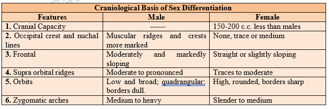
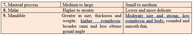
Determination of Sex
A sexual diagnosis from fragmentary, isolated remains without possibility of comparison is extremely difficult as in case of fossils hominids. Estimation of sex in complete human skeleton is usually easy provided it is post pubertal. Sexual differences are marked in pelvis and skull, but not equally in all populations. To diagnose sex from a skull is a rather difficult task because a female skull can show exceptional masculine characters. Similarly, the male skull can also show feminine characters. Moreover, the sexual differences between the skulls may be race specific. Sex determination may be easier in Whites than in Negroids or Mongoloids.
A number of features (descriptive and metric characters) must be considered together. The co-existence of several features may permit a judgement.
Facial Superimposition
This anatomic material can be used for identification when the skull and facial bones are used as a foundation for the reconstruction of facial soft tissues. With the use of standard anthropological thickness measurements at specific points on the face, soft-tissue thickness points can be connected with sculpting clay and the reconstructed features can sometimes be digitized on a computer screen. Because computers permit the addition of components directly to cranial features, computers have been useful for techniques involving facial superimposition. The underlying skeletal structures can thus be viewed below the soft tissue, providing a means to check its accuracy. The result of these techniques is a recreation of the contour of the soft-tissue features that permits visual identification. Various versions can then be stored and reproduced for comparison.
Method of Identification from the Suture Pattern
An effective method of identification from the suture pattern found on the ectocranial surface of skull; means physical pattern evidence which shows that each skull possesses a unique configuration of suture formation. The suture spikes, projection, denticulate, recess, and other type of irregularities, are so varied that they are thought to be as individualistic as finger ridge.
In order to have confidence in the use of suture patterns for personal identification the following three basic principles must be established,
1. Individual skull suture patterns should be so highly variable that their characteristic even in a subdivision of suture, are not duplicated elsewhere in the suture, in other sutures or in a different individual.
2. The configuration and details of individual suture patterns should be permanent.
3. The configuration types should individually be variable but varying within limits which permit systematic classification.
Nevertheless, the personal identification of the dead as well as the living through surface configuration formed on the ectocranial surface is a violable system which may assume greater importance in the near future.
Uniqueness of Suture
Evaluation of suture patterns shows that the skull suture pattern configurations are unique. The frequencies of patterns in both the sexes have been found to be more or less equal and hence the attribution of sex to skull from suture patterns and their configuration is questionable. Suture configurations are found to be not bilaterally symmetrical. The bilateral differences exhibited by the pattern configuration, that suture pattern in a skull are highly individualistic, and the patternings are not duplicated in another segment of the same suture.
Permanence of Suture Pattern
Suture patterns can only be used as a means of identifying a person, if the premortem pattern is recorded radiographically; although lateral and oblique views may contain suture lines, the antero– posterior view is most suitable for radiographically recording the sutures for the purposes of personal identification. In older individuals the number of segment recorded radiographically is found to decrease gradually with age progression.
Systematic Classification of Suture Pattern
A system of classification of suture patterns has been evolved by gathering the patterns into the following ten classes:
· Serrate type of suture
· Denticulate type of suture
· Serrated dentate type of suture
· Dentated serrate type suture
· Wavy type of suture
· Crenate type of suture
· Undulate type of suture
· Closed type of suture
· Corolla type of suture
· Complicated type of suture
The ectocranial suture patterns in them are highly individualistic and that no two skulls can ever have an identical pattern. A positive method of identification of the skull is suggested by the comparison of radiographic with visible skull suture pattern. Positive identification through radiographic comparison of antemortem and postmortem cranial suture patterns evaluated in light of these criteria. Computed tomography (CT) scans may prove a more useful modality for positive identification, due to better resolution and greater availability. The skull vault is a dynamic structure formed from multiple membranous flat bones connected at the edges by fibrous sutures. As the brain grows during development, free expansion of the cranium occurs at the sutures.
Summary
Bones are live and carry all life functions. The condition of bones can tell investigator about a person’s health and nutrition during life. The identification of factors differentially expressed in fusing sutures has led to multiple targets for future research. Evaluation of suture patterns shows that the skull suture pattern configurations are unique. Individual skull suture patterns should be so highly variable that their characteristic even in a subdivision of suture, are not duplicated elsewhere in the suture, in other sutures or in a different individual.
The skull suture patterns have been suggested for use in individualizing human remains by comparing antemortem and postmortem radiographs. The suture pattern on skulls remains constant throughout the life time of an individual. An advantage in the process of identification through suture pattern is that they cannot easily be altered or destroyed even in cremated remains. Suture patterns can only be used as a means of identifying a person if the premortem pattern is recorded radiographically. A number of features (descriptive and metric characters) must be considered together. The co-existence of several features may permit a judgment.
| you can view video on Skull suture pattern individualization based on suture patterns ANT |