11 Microscopic and gross anatomy of human bones

Introduction
The body is shaped and supported by a framework of bones called the skeleton; which protect internal organs, such as the brain, and lungs, and provide an anchorage for the muscles. Bones are live and carry on all life functions. The condition of bones can tell investigators about a person’s health and nutrition during life. Bones develop from cells known as osteoblasts, first beginning as soft cartilage before the bone hardens through the introduction of various minerals, a process known as ossification.
Skeletal System Function: Support and protection and assistance in movement
Bone is one of the hardest structures of the animal body; it possesses also a certain degree of toughness and elasticity. Its colour, in a fresh state, is pinkish-white externally, and deep red within. The human body bones that perform five main functions:
- Support
- Protection
- Body movement
- Blood cell formation
- Storage of inorganic salts and lipid
Skeletal System Function: Mineral homeostasis, blood cell production, triglyceride storage
- Support for soft tissue and provision of attachments for many skeletal muscles.
- Protection for the brain and spinal cord, and thoracic organs.
- Movement – bones used as levers for skeletal muscles. Joints allow movement.
- Storage and release of minerals especially calcium and phosphorus.
- Storage of fat as an energy reserve in yellow marrow.
- Blood cell formation – most haematopoiesis (blood cell formation) occurs in the marrow cavities of certain bones.

Bone
Bones are made of a hard, living, self-repairing tissue that is supplied with blood vessels and nerves. Bone consists of widely spaced osteocytes (bone cells) and the matrix that lies between them. The matrix is made up of fibres of collagen, which give bone its flexibility, and mineral salts, mainly calcium phosphate, which gives bone its strength. Surrounding all bone is a layer of hard, compact bone. The spaces within spongy bone are frequently filled with red marrow. It is important for bones to be strong to support our body weight and in some cases provide protection such as the skull and ribs. However, they must also be light enough to make movement possible.
Classification of Bone Shape
Bones can be divided into a number of classes;
- Short,
- Long,
- Flat,
- Sesamoid and
- Irregular bones.
Short bones, such as the carpal bones within the wrist, tend to be as wide as they are long. Long bones are, as the name suggests, longer in length and also tend to be slightly curved, for example the femur. Flat bones, such as the ribs and breastbone, could be described as being fairly flat and plate-like. Sesamoid bones refer to small bones embedded in a tendon, often found in joints, such as in the knees and wrists. Finally, irregular bones refer to a certain class of bone that do not belong in the other categories, such as the bones composing the spine.
Types of Bone
A. Lamellar or compact bone
- Contains osteons composed of concentric lamellae
- Each Osteon has an Osteonic canal (Haversian canal) which has blood vessels and nerves
- Osteocytes found within lacunae
- Canaliculi connect osteocytes and function to diffuse nutrition to the osteocytes
- Communicating (also called perforating or Volkmann’s) canals, connect adjacent osteons, and carry blood vessels.
- The diaphysis, or shafts of long bones, are composed mostly of lamellar bone
B. Cancellous, trabecular, or spongy bone
- Does not contain Haversian canal systems (osteons)
- Nerves and blood vessels run randomly through the loose meshwork of bone.
Bone Shape and Gross Appearance
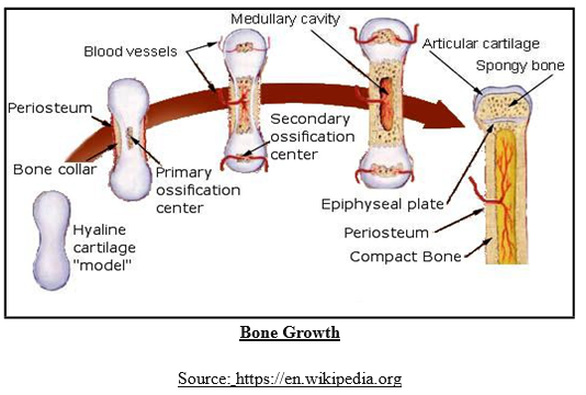
Bones vary in their shape and gross appearance. Their shape and structure is affected by genetic, metabolic and mechanical factors. Each bone is the result of a long functional history through innumerable successive generation. The fact that the primary shape of the bone is genetically determined is well demonstrated by transplantation of embryonic skeletal tissues and organ culture studies. Other characteristics of the bone seem to be self determined. Mechanical influences may not be operative at the time of establishment of the primary form of the bone. Later on muscles become active and may influence bone growth even in the pre- natal life. There is a lot of increased activity which augments growth both in length and circumference till the epiphyses are fused.
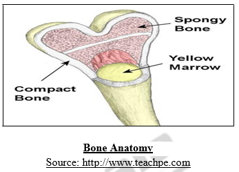
Bone Anatomy
Metabolic influences effect bone growth at all stages of development. It is known that osteogenesis necessarily requires the availability of calcium, phosphorous and Vitamin – A, C, D along with the secretions of the hypophysis, thyroid, parathyroid, adrenal, glands and gonads. All these processes are therefore essential for skeletal form and dimensions. If we were to cut a cross-section through a bone, we would first come across a thin layer of dense connective tissue known as Periosteum. This can be divided into two layers, an outer ‘fibrous layer’ containing mainly fibroblasts and an inner ‘cambium layer’, containing progenitor cells which develop into osteoblasts (the cells responsible for bone formation). The periosteum provides a good blood supply to the bone and a point for muscular attachment.
Under the periosteum is a thin layer of compact bone (often called cortical bone), which provides the bones strength. It consists of tightly stacked layers of bone which appear to form a solid section, although do contain osteons, which like canals provide passageways through the hard bone matrix.
Bone Marrow
Bone marrow is a jelly-like material found inside bones. Red bone marrow inside the hip, skull, collar bones, sternum, and backbone is the site of blood cell production. Red marrow is the site of red and white cell production.
Spongy Bone
Spongy bone is a “honeycomb” layer that lies beneath compact bone. It forms a light but strong framework that reduces the bone’s weight but not its strength. The other consists of slender fibers and lamellae, which join to form a reticular structure; this, from its resemblance to lattice-work, is called spongious tissue. The compact tissue is always placed on the exterior of the bone, the cancellous in the interior. The relative quantity of these two kinds of tissue varies in different bones, and in different parts of the same bone, according as strength or lightness is requisite. Close examination of the compact tissue shows it to be extremely porous, so that the difference in structure between it and the cancellous tissue depends merely upon the different amount of solid matter, and the size and number of spaces in each; the cavities are small in the compact tissue and the solid matter between them abundant, while in the cancellous tissue the spaces are large and the solid matter is in smaller quantity. Struts link to form a framework.
Combat Bone
Bone Anatomy
Compact bone forms the outer part of bone. After teeth enamel, compact bone is the hardest material in the body. It is made up of parallel cylinders called Osteons. Each Osteon consists of layers of lamellae. Bone looks dense and solid, but full of passageways that serve as conduits for nerves, blood vessels, and lymphatic vessels.
Blood and nerve supply of bone
Blood is supplied to mature compact bone through the Haversian canal, formed when individual lamellae form concentric rings around larger longitudinal canals within the bone tissue. These Haversian canals typically run parallel to the surface and along the long axis of the bone. The canals and the surrounding lamellae are called a Haversian system or an osteon. A Haversian canal generally contains one or two capillaries and nerve fibers. The Haversian canals also surround nerve cells throughout the bone and communicate with osteocytes in lacunae (spaces within the dense bone matrix that contain the living bone cells) through canaliculi. This unique arrangement is conducive to mineral salt deposits and storage which gives bone tissue its strength.
Bone Formation
Major cell types in bone tissues
- Osteogenic,
- Osteoblasts,
- Osteocytes,
- Osteoclasts
Structure and Development of Bone
The bones and teeth of the craniofacial complex, key identification tools for the forensic odontologist, effectively distinguish one person from others and one population from another and are used to determine the race, age and sex of a person. The adult human skeleton consists of some 206 individual bones, with there being even more in the skeleton of a child, whose bones have not undergone certain fusion processes yet, and many of these bones may prove useful to the anthropologist.
Levels of bone structure
- Gross,
- Microscopic, and
- Chemical
Bone during life is permeated by vessels, and is enclosed, except where it is coated with articular cartilage, in a fibrous membrane, the periosteum, by means of which many of these vessels reach the hard tissue. If the periosteum be stripped from the surface of the living bone, small bleeding points are seen which mark the entrance of the periosteal vessels; and on section during life every part of the bone exudes blood from the minute vessels which ramify in it.
Histology of Bone Tissue
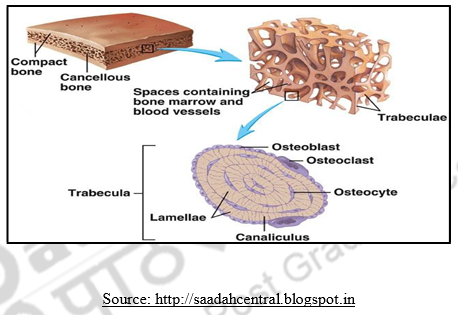
- Lamella: Lamella is one of the tubes of bone surrounding a Haversian canal.
- Lacuna: Lacuna is a space that contains osteocytes (bone cell).
- Haversian canal: Haversian canal is a space that runs down the centre of the Osteon, carrying blood vessels and nerves.
- Blood vessels: Blood vessels supplies bone cells with oxygen and food.
- Osteon: An Osteon is a small piece of compact bone made up of tiny tubes called lamellae arranged in circular layers around a central Haversian canal.
Transverse Section of Dense Bone
Periosteum
The periosteum adheres to the surface of each of the bones in nearly every part, but not to cartilaginous extremities. When strong tendons or ligaments are attached to a bone, the periosteum is incorporated with them. It consists of two layers closely united together, the outer one formed chiefly of connective tissue, containing occasionally a few fat cells; the inner one, of elastic fibers of the finer kind, forming dense membranous networks, which again can be separated into several layers. In young bones the periosteum is thick and very vascular, and is intimately connected at either end of the bone with the epiphysial cartilage, but less closely with the body of the bone, from which it is separated by a layer of soft tissue, containing a number of granular corpuscles or osteoblasts, by which ossification proceeds on the exterior of the young bone.
Later in life the periosteum is thinner and less vascular, and the osteoblasts are converted into an epithelioid layer on the deep surface of the periosteum. The periosteum serves as a nidus for the ramification of the vessels previous to their distribution in the bone; hence the liability of bone to exfoliation or necrosis when denuded of this membrane by injury or disease. Fine nerves and lymphatics, which generally accompany the arteries, may also be demonstrated in the periosteum.
A transverse section of dense bone may be cut with a saw and ground down until it is sufficiently thin. If this be examined with a rather low power the bone will be seen to be mapped out into a number of circular districts each consisting of a central hole surrounded by a number of concentric rings. These districts are termed Haversian systems; the central hole is Haversian canal, and the rings are layers of bony tissue arranged concentrically around the central canal, and termed lamellae. Moreover, on closer examination it will be found that between these lamellae, and therefore also arranged concentrically around the central canal, are a number of little dark spots, the lacunae, and that these lacunae are connected with each other and with the central Haversian canal by a number of fine dark lines, which radiate like the spokes of a wheel and are called canaliculi.
The Lacunae are situated between the lamellae, and consist of a number of oblong spaces. In an ordinary microscopic section, viewed by transmitted light, they appear as fusiform opaque spots. Each lacuna is occupied during life by a branched cell, termed a bone–cell orbone–corpuscle, the processes from which extend into the canaliculi.
Canaliculi
The Canaliculi are exceedingly minute channels, crossing the lamellae and connecting the lacunae with neighbouring lacunae and also with the Haversian canal. From the Haversian canal a number of canaliculi are given off, which radiate from it, and open into the first set of lacunae between the first and second lamellae. From these lacunae a second set of canaliculi is given off; these run outward to the next series of lacunae, and so on until the periphery of the Haversian system is reached; here the canaliculi given off from the last series of lacunae do not communicate with the lacunae of neighbouring Haversian systems, but after passing outward for a short distance form loops and return to their own lacunae. Thus every part of an Haversian system is supplied with nutrient fluids derived from the vessels in the Haversian canal and distributed through the canaliculi and lacunae.
The bone cells are contained in the lacunae, which, however, they do not completely fill. They are flattened nucleated branched cells, homologous with those of connective tissue; the branches, especially in young bones, pass into the canaliculi from the lacunae.

Cartilage
When we were born, our bones were not as strong and hard as they are now. This is because when we were babies, our bones were mostly made of cartilage. A cartilage is the soft flexible substance that we can feel when we press our ears and nose. As we grow, the cartilage slowly transforms into dense and hard bones. Our bones stop growing after we reach the age of 25. However, a point to be noted is that not all cartilage transform into bones. Apart from your ears and nose, cartilage can be found in places including the joints of the bones, ribcage, bronchial tubes, and intervertebral discs. This enables smooth movements between the bones since the cartilage protects them from rubbing against each other.
Bone Fractures
A fracture, or break, happens when a bone is exposed to a sudden force that it cannot withstand. There are two types of fracture. In simple or closed fracture, the broken bone ends remain below the skin; whereas in compound or open fractures, they stick out through the skin and often cause damage to surrounding tissue. Fractures bones mend themselves.
Types of Fractures
Fractures may be classified by position, completeness of the break, orientation of the break relative to the long axis of the bone, and whether the bone ends penetrate the skin or not.
- Comminuted,
- Compression,
- Spiral,
- Epiphyseal,
- Depressed,
- Greenstick
Remodeling of Bone
Bone remodeling occurs starting during bony callus formation and continuing for several months after, the bony callus is remodeled. The excess material on the diaphysis exterior and within the medullary cavity is removed and compact bone is laid down to reconstruct the shaft walls. the final structure of the remodeled area resembles that of the original unbroken bony region because it responds to the same set of mechanical stressors.
Factors affecting bone growth and remodeling Minerals
(a) A blood clot forms where bone is broken.
(b) New blood vessels forms between broken ends
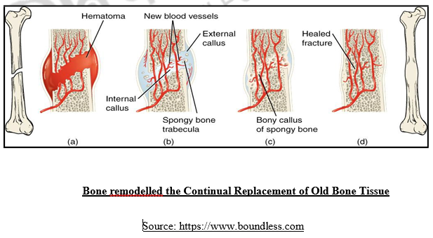
(c) Spongy bones form between broken ends.
(d) Fracture has healed and bones returns to its original shape.
The osteoclasts identify that the bone has reached the end of its life cycle and break down its tissues. The osteoblasts then, make sure that new bone tissues take place of the old ones, making sure that the bones are always as good as “new”
Imaging bones
Doctors use X-rays to look inside the patient’s body for sign of damage or disease, without surgery. X- rays are type of radiation that passes through the body’s soft tissue but not through bone. An X-ray machine produces a negative called a radiograph, in which only the bone shows up body.
Osteoporosis
Osteoporosis refers to a group of diseases in which bone resorption outpaces bone deposit; bone becomes so fragile; bone mass is reduced; spongy bone is most vulnerable; osteoporosis occurs most often in middle aged, and post menopausal women.
Factors that Contribute to Osteoporosis
- petite body,
- insufficient exercise,
- immobility,
- a poor diet in calcium and protein,
- smoking,
- abnormal vitamin D,
- hormone related conditions
Reducing the risk of osteoporosis
Rich source of calcium, a vital ingredient for strong bones and reducing the risk of osteoporosis later in line. Together with another mineral called Phosphate; it provides main strength in bones but also helps to increase the power of muscles. Getting enough calcium in the diet during childhood is crucial for strong bone. Osteomalacia and rickets are both caused by insufficient calcium in the diet or by a vitamin D deficiency.

Summary
Bones are dynamic structures that are undergoing constant change and remodeling in response to the ever-changing environment. In fact, there is so many turnovers that in 4 years, the skeleton of a young person will be completely new as compared with their skeleton today. Bones can react and respond to environmental stimuli; they can get bigger or smaller, they can strengthen themselves when needed, and, when broken, they are among the few organs with the ability to regenerate without scar. Bones of the skeletal system surround and protect organs, such as the brain and heart. Human bones are very strong and can resist tremendous bending and compression forces without breaking.
The is allows the major features of individual bones to be seen clearly without being obstructed by associated soft tissues, such as muscles, tendons, ligaments, cartilage, nerves, and blood vessels. The important relationships among bones and soft tissues should not be ignored. Automatic detection and treatment is given utmost priority in medical field. To develop an efficient automatic bone fracture detection and identification system, a clear understanding about the human skeletal system and fractures is required.
| you can view video on Microscopic and gross anatomy of human bones |