31 Cytogenetic, Immunogenetics and DNA methods to solve the Paternity Dispute Cases
1. Introduction:
DNA evidence is used in court to prove the paternity dispute cases, rapes, criminal cases. In paternity cases, if particular man has allele which is strongly matching with child’s allele, at certain locus, he cannot be excluded from the paternity dispute. In some cases it also seems that close relatives will be more likely to have the child’s paternal allele. Forensic genetics has focussed on the Y-STR markers, which are available for population genetics, and forensic investigation. In this chapter discussion about the paternity dispute cases, how cytogentics is useful to determining certain cases related to forensic investigation and the methods of the DNA extraction are discussed. The rapid development in the technology such as STR typing technology, has increased power to identify real fathers thus providing the objective evidence for solving civil and criminal cases.
2. Cytogenetic :
It is the study of the cell chromosome and their role in hereditary structure. The term chromosome was coined by Walder in 1888. Several discoveries were made to correct the numbers of the chromosomes. In 1955 two scientists in Sweden, Lund and Tijo were experimenting with the culture of embryonic lung cells established 46 correct numbers of chromosomes. This study was confirmed by other scientist Ford and JA Hammerton on studies on human spermatocytes. Chromosomes are 46 in number. Out of these 23 pairs, 22 pairs constitute the autosomes and are similar in males and females. 23 rd pair of chromosomes are known as sex chromosome. Female chromosomes are XX and males having XY chromosomes in the 23rd pair of chromosomes. Later in 1960 a group of scientists working in Denver in Colorado in U.S.A. adopted a system for classifying and identifying the human chromosomes according to their length, positions of centromere as metacentric, submetacentric and acrocentric. The chromosomes are placed according to their length. The longest chromosomes are placed at starting of the order of sequences and shortest at the end. This system is known as Denver System.
2.1. Chromosomes are divided into seven groups.
According to the numbering order placed.
- Group A. Chromosomes pairs include from 1-3. Here chromosomes are grouped according to in the position of centromere and by the length of chromosomes. This group contains three largest chromosomes.
- Group B. Chromosome from 4-5. Chromosome 5 is slightly smaller than chromosome 4. According to the positions of the sub terminal
- Group C. Chromosome from 6-12. Chromosome 6, 7, 8, and 11 are metacentric. While 9, 10, 11 are at sub terminal.
- Group D. Chromosome from 13-15.
- Group E. Chromosome from 16-18.
- Group F. Chromosome from 19-20.
- Group G. Chromosome from 21-22.
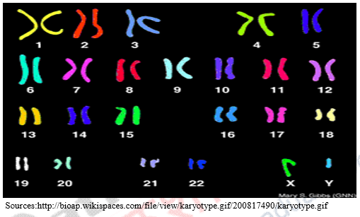
From figure given below chromosome groups can be understood. Chromosome 1-3 group as Group A, 4-5 group B, 6-12 group C, 13-15 Group D, 16-18 Group E, 19-20 Group F, 21-22 group G. And lastly sex chromosomes which are numbered as 23rd chromosome. It can also be seen clearly that length of chromosomes are arranged in from largest to shortest end.
In moderns techniques chromosomes are stained and have different banding patterns and arranged into standardized format. This standardized format is called karyotype.
2.3. Banding Patterns in Human Chromosome:
The technique used by staining the chromosome in metaphases for cations identified is called banding. A band is defined as that part of chromosome which is clearly distinguished from the adjacent segments by appearing darker or brighter with one or more banding techniques. Banding techniques are having two principle groups. I). The band are distributed with the length of chromosome such as G band, Q band, R Band. 2) Those that stains a restricted number of specific bands or structure are named C band.
There are four banding patterns, as follows:
- Q Band (stain Quinacrine);
- G band (stain: Giemsa): it named after Giemsa dye. In G band, dark regions tend to be hetrochromatic.
- R band (reverse of Giemsa). Its reverse of G band. The dark regions are euchromatic and bright regions are heterochromatic.
- C band (constitutive heterochromatin).
2.4. Abnormal Chromosomes:
Human chromosomes are 46 in numbers. Chromosome abnormalities are study based on autosomal aberrations and sex-chromosomal aberrations.
Autosomal aberrations include number and structure of autosomal chromosomes. Autosome means (Autovysome body chromosomes). Numerical autosomal include aneuploidy and polyploidy.
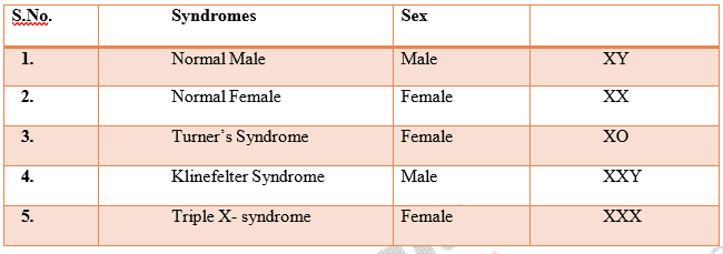
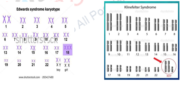
2.5. Sex Chromosomes Abnormalities:
2.6. Chromosomal Aberrations
2.6 Paternity Test
1. Y chromosome in for forensic analysis for paternity test: Y chromosome is very useful in determining the paternity. Y haplotyping application for special instances, such as deficiency case in paternity testing and analysis of mixtures of male and female DNA or with combination with autosomal markers. It is useful in the cases like violent crimes done by the males, sexual offence. So at crime scene culprits’ leaves samples usually informative of the Y markers in rape cases. In the case of paternity of son is in question so in this case comparison of the Y chromosome will exclude the alleged father.
2.6.1. Features of the Y chromosomes: Y chromosomes are found only in male sex. And it passes from father to son. Y chromosome is paternally inherited and haploid. Its haploid so it cannot recombine with other at meiosis. it can be done by using DNA polymorphism.
1. Y polymorphsism and Y chromosome tree
a). Binary polymorphism.
b). Micro Satellite and mini satellites.
c). Haplotypic diversity.
2.6.2. Practical applications of Y Markers: They are using small PCR amplicons. Y chromosome is suited for analysis of DNA samples. Characteristic of PCR is that it is large cluster of repeat units.
Cases: Y chromosome analysis for paternity testing. In this case alleged father was not available. Some of relatives, paternal uncle of putative son were available. These people were taken for Y chromosome marker test. Exclusion can be done on the basis, if the Y chromosome is not same as putative son.
Case 2. Rape case: semen of attacker is mixed with victims cells. Then sperm nuclei of attacker are separated from the component of female victim by the differential lysis of cell types. If in the case this method doesn’t give the results, then victim female profile is separated from the attackers profile and the autosomal marker is used for analysis isused. On the other hand in such cases of rape Y-specific analysis allows attacker’s haplotype to be determined. In cases like multiple rapes, microsatellite of Y specific allows to determined the numbers of attackers. Analysis of haplotypes of detained suspects could show whether the mixed haplotype were consistent with match or not. Y- chromosomal markers will be very useful in analysis of other kind of mixtures blood-saliva, blood-blood. Here differential lysis cannot be apllied. Y- chromosome haplotype could give officers of police, the surname of the suspects who left evidence at crime place. Because in many societies surname are co- inherited so Y – chromosome are co-inherited with surname.
3. DNA (Deoxyribo nucleic Acid)
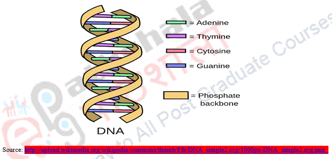
DNA is basically made up of long molecules that contain coded instructions for the cells. DNA contains long molecules chains. The link of the chain is in pieces and called nucleotides also called bases. There are four different types of nucleotides called A, G, C, T. The back bone of DNA is made up of phosphate.
3.1. The techniques that are used for identifying testing. They are following:
- DNA Profiling.
- DNA Finger Printing.
- DNA Typing.
3.2. History of DNA Finger Printing:
In the beginning, the identification of a person was done through the ABO blood group system. Later new markers were discovered on the basis of the protein serum and red blood cells enzymes. Later Sir Alec Jeffery, invented DNA finger printing techniques.
3.3. About DNA Finger Printing:
- It’s technique to determine sequence of nucleotide in certain area of DNA. The sequences of nucleotides are unique to individual person.
- Every person has unique fingerprinting.
- DNA fingerprints are same for every cells, tissues and organs of the individuals.
- Fingerprints cannot be changed throughout the life time.
- Individual person can be distinguished by the entire genomic DNA sequences.
3.4. Principle of DNA Finger Printing
Human genome consists of several small non coding and inheritable sequences of the bases. These sequences of bases are repeated several times. The sequence occurs near Y chromosome, hetrochromatin area, centromere, telomere. The area where sequence of DNA is repeated many times are called repeated DNA. Repeated DNA is separated as satellite from the bulk of DNA by centrifugation, density gradient and this is called Satellite DNA. The repetition of the basis of satellite DNA is called Tandem. Satellite DNA shows the polymorphism. Short nucleotides repeat since the DNA are very specific in each individual and vary in number from person to person and is called VNTR (Variable numbers tandem repeat). These are also known as “Minisatellite”. This is used as genetic markers for each individual personal identity.
3.5. Techniques Used for DNA Finger
1. First DNA is extracted from the White blood cells, spermatozoa, skin and from the hair. Hair that is forcefully pulled out or natural fallen. From the roots of the hair DNA is extracted.
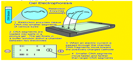
2. Then DNA molecules are cut into fragments by the chemical knife with the help of enzyme restrictions endo nucleus. This fragment of the DNA also contains the VNTRs.
3. On the Gel electrophoresis the fragments are separated according to the size.
4. Particular size of the fragments contains VNTRs which multiply by the PCR (polymerase chain reaction) technique. To split them into single strand of the DNA they are treated with the alkaline chemicals.
5. Single strands of DNA are transferred to the nylon membrane.
6. On the nylon membrane radioactive probes of the DNA consisting the possible VNTRs are put. then nylon membrane are washed to extra probes. This process involves the Southern blotting technique. It is named after the inventor E.M. Southern. The method of hybridization of DNA with the probes is called Southern blotting.
7. On nylon membrane X-ray film are exposed to probes of the DNA to mark the places where radioactive probes of DNA are bound to DNA fragments. The places are marked as dark band after X-ray film is developed. This technique is known as autoradography.
8. On the X-ray films the dark band represents the DNA fingerprinting.(DNA Profile).
3.6. Application of DNA Fingerprinting:
- Identifying the human remains.
- Paternity and Maternity dispute.
- Studying the relation with ancient humans.
- Accused and convicted fellows can be set free because of DNA typing.
- Useful in detection of the crime and legal pursuits.
- Determining the bone marrow transplants.
- Proving the relatedness of immigrations.
- Lineage markers.
- Hereditary disease.
- Finger printing can help individuality of uniqueness.
3.7. Paternity Determinations:
1) Paternity determination through allele frequency distribution of highly polymorphic DNA sequences.
In this determinations of paternity blood samples are collected from paternity cases. Sample types for Blood group, HLA antigens and series of protein polymorphism, for the inclusion of the paternity probability of alleged father are calculated using allele frequency. Alleged father is excluded on the basis of the size of his alleles if they are different from the inherited alleles of the child’s paternity. This is done by taking each individual’s phenotype alleles. Blood samples are collected. Then cases of paternity samples of DNA are digested with EcoRI or TaQI and loaded into agarose gels for fractionation. If there are no detectable difference between one of the alleles of the alleged father and child then there is the probability of his being biological father of the child.
3.7.1. Applications of DNA polymorphism for the determinations are the following:
a) Blood samples taken from paternity case are studied by protein ( RBC protein and HLA typing).
b) DNA fragments are cut by the enzymes restricted endonuclease.
c) Fragments of the DNA are done by the agarose gel electrophoresis hybridized with radioactively labelled DNA probes.
d) Finally the size of the allele detected by these two probes.
3.7.2. Paternity Testing with locus incompatibility:
A case study of SNPSTR efficiency in paternity testing with locus incompatibility methods used for this test is: Based on the short tandem repeats (STR) loci analysis to observe the more common incompatible STR locus.
a) DNA samples from the paternity cases were extracted.
b) Then Single nucleotide proteins and CSF1PO genotyping.
c) Single nucleotide protein (SNP), short tandem repeats (STR) haplotype analysis. d). DNA sequencing.
e) finally done with statistically analysis.
Here observation for paternity test is that SNPSTR can be applied for the paternity testing. The power of SNPSTR are much greater than the alone STR and SNP alone. SNP are also useful and determine to distinguish fathers from uncles.
Case study: Determination of paternity from aborted, chronic villi: the paternity test is more difficult for aborted child. Maternal DNA is ample then the DNA of foetal and its preferred to replicate in the PCR (polymerase chain reaction) analysis. A case of mentally retarded and physically disabled women 21 years old was taken to emergency room of the hospital because she was suffering from the vaginal bleeding. Doctors found during examination that she passed a large clot of blood from vaginal bleeding. By histological examination it was found that she had suffered from abortion at early stage of pregnancy. Because from histological examination the presence of the chronic villi, was known. Chronic villi give genetic material to chronic villus cells in the foetus. Its like finger shape and very small in size. It is formed by the embryo by extra embryonic tissue layers. And thus becomes the part of the uterus that victims shed after birth. After histological analysis doctors reported it to the criminal investigation case. As victims was unable to give consent to sex. So it was concluded that she was sexually assaulted. Victims conditions also show that she was unable to recognised who had assaulted her. So only way left to find out the culprit was to do DNA test. Blood sample from the women was taken. To know the culprit laser micro dissection to isolate tissue from foetal chronic villi was done without including surrounding maternal tissues. Collected samples from victims and suspect were analysed by short tandem repeats (STR) for 15 loci and amelogenin locus for sex determination. It was found that foetal aborted was of female sex and in the profile of foetal tissue isolated, one allele is common to the victim women, indicating that she is the mother. Another 8 loci in the foetus were homozygous and it strongly indicated that women were assaulted by someone closely and genetically related to her. So to know the culprit, samples were taken from her brother, father, uncle, maternal and paternal both grandfathers. Samples was again analysed on the STR (short repeat tandem). The result shows that allele was incompatible with the maternal, paternal grandfather and uncle and with the father. So they were excluded as culprit suspects. However victims women’s brother showed the strong match with each of 15 loci on the foetus profile of STR and probability showed he was responsible.
Case of Paternity dispute from Hyderabad: the husband’s plea is that he had no access to the wife when the child was begotten can be proved by the DNA test report and in the face of it, court was not able to decide the fatherhood of a child. Then court decided to send them for the DNA test for scientific results. When the scientific reports proved to the contrary and further the innocent child may not be bastardized as the burden is on the mother to prove that the revision petitioner is the father, from the marriage also in dispute, equally of any conjugal life.” While dismissing the revision petition, justice Siva Sankara Rao ruled that the DNA test helps as a solid proof on truth of the dispute of paternity.
4. Immunogenetics:
Ability to induce an antibody or cell mediated response. Where an antigens substance which can be recognised by the surface antibody (B cells) is associated with major histocompatibility molecules. HLA genes and Major histocompatibility is activating and inhibitory killer immunoglobulin like receptors gene from haplotype, which shows diversity not only reflects in only nucleotides difference of the alleles, but also in terms of presence or absence in individual genes.
HLA is use to match a donor of bone marrow, blood transplant. HLA is protein found in all cells of the body. The immune systems uses HLA as markers to know which cells belong to one individual’s body and not.
Different donor features have impact on transplant success. They include: Age, Gender, Blood type and the number of time the female donor has been pregnant. The method used to test HLA antigens and antibodies early are based on the serological methods. Now antigens are molecularly defined by the DNA analysis such as flow cytometry, solid phase immunoassay and single antigen bead assay such as Luminex.
In recent years to more advance techniques have been developed to determine the paternity i.e.
1. Human Leukocyte Antigen (HLA): High degree of discrimination of HLA systems make more accurate probability to determine paternity. It caused the revolution in paternity determination after combining HLA with red cells antigens test.
Case 1. For HLA: The New York court generally accepted the HLA for paternity dispute solving in the case of Goodrich V. Norman. Court held that alleged father has right to not exclude from the HLA red cells antigen test. Judge of the court found HLA techniques are widely accepted by the scientist. It proved that organ transplant is used to match the donor and recipients. It’s very important when dealing with the lives of the patients.
Case 2. For HLA: In New Jersey Malvasi vs. Malvasi case. Court granted the putative father motions to oblige mother to have HLA testing. Because HLA testing was highly recognised by the scientific community.
- Red cell enzymes and blood serum protein by electrophoresis method: Many Blood serum proteins and red cell enzymes have proven useful in determining the paternity test by electrophoresis method. Because proteins and enzymes process follow the Mendelian inheritance patterns.
5. Summary:
DNA extraction from biological materials such blood group systems, protein of blood, red cell antigens, Haplotype, SNP and STR are very useful in determining the case such as rape cases, paternity disputes inclusion and exclusion of the alleged father. Most of the paternity test is done for the financial reasons to get the legal responsibility of the legal support to the child. Such test can determine the important tools for civil court, when paternity of a child is question. The methods described in this chapter are STR, SNP, allele frequency compatibility. Which don’t include for unrelated, they can be applied for the consideration of genetic relationship. Serological test for identifying a person is now a old trend. So HLA typing is good to identify a person.
| you can view video on Cytogenetic, Immunogenetics and DNA methods to solve the Paternity Dispute Cases |


